

Single-chain recombinant HLA-DQ2.5/peptide molecules block α2-gliadin-specific pathogenic CD4+ T-cell proliferation and attenuate production of inflammatory cytokines: a potential therapy for celiac disease
- PMID: 20736999
- PMCID: PMC3012747
- DOI: 10.1038/mi.2010.44
Single-chain recombinant HLA-DQ2.5/peptide molecules block α2-gliadin-specific pathogenic CD4+ T-cell proliferation and attenuate production of inflammatory cytokines: a potential therapy for celiac disease
Abstract
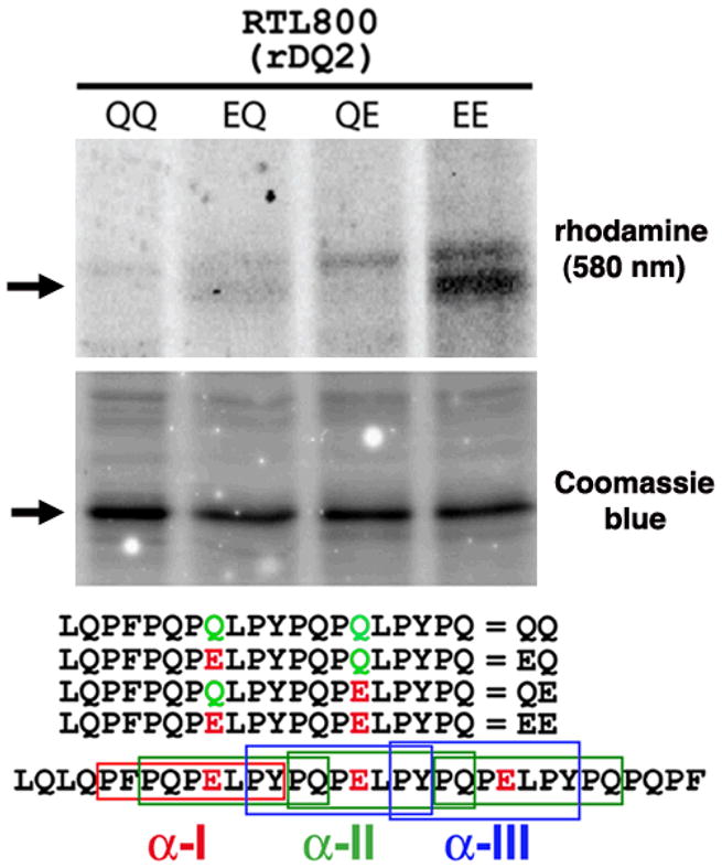
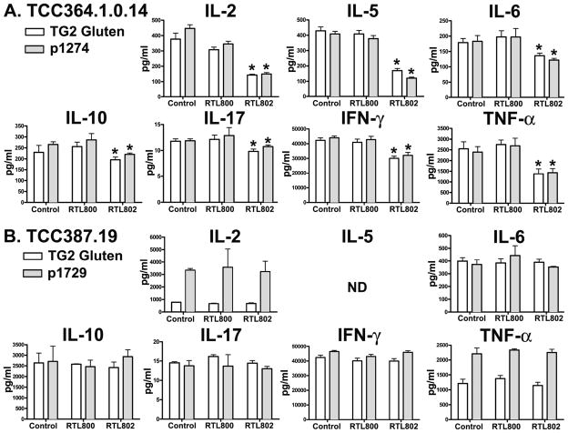
Celiac disease (CD) is a disorder of the small intestine caused by intolerance to wheat gluten and related proteins in barley and rye. CD4(+) T cells have a central role in CD, recognizing and binding complexes of HLA-DQ2.5 bearing gluten peptides that have survived digestion and that are deamidated by tissue transglutaminase (TG2), propagating a cascade of inflammatory processes that damage and eventually destroy the villous tissue structures of the small intestine. In this study, we present data showing that recombinant DQ2.5-derived molecules bearing covalently tethered α2-gliadin-61-71 peptide have a remarkable ability to block antigen-specific T-cell proliferation and inhibited proinflammatory cytokine secretion in human DQ2.5-restricted α2-gliadin-specific T-cell clones obtained from patients with CD. The results from our in vitro studies suggest that HLA-DQ2.5-derived molecules could significantly inhibit and perhaps reverse the intestinal pathology caused by T-cell-mediated inflammation and the associated production of proinflammatory cytokines.
Conflict of interest statement
Figures


Similar articles
-
The intestinal T cell response to alpha-gliadin in adult celiac disease is focused on a single deamidated glutamine targeted by tissue transglutaminase.J Exp Med. 2000 Feb 21;191(4):603-12. doi: 10.1084/jem.191.4.603. J Exp Med. 2000. PMID: 10684852 Free PMC article.
-
Antigen presentation to celiac lesion-derived T cells of a 33-mer gliadin peptide naturally formed by gastrointestinal digestion.J Immunol. 2004 Aug 1;173(3):1757-62. doi: 10.4049/jimmunol.173.3.1757. J Immunol. 2004. PMID: 15265905
-
Resistance to celiac disease in humanized HLA-DR3-DQ2-transgenic mice expressing specific anti-gliadin CD4+ T cells.J Immunol. 2009 Jun 15;182(12):7440-50. doi: 10.4049/jimmunol.0900233. J Immunol. 2009. PMID: 19494267
-
Transglutaminase 2 and Transglutaminase 2 Autoantibodies in Celiac Disease: a Review.Clin Rev Allergy Immunol. 2019 Aug;57(1):23-38. doi: 10.1007/s12016-016-8557-4. Clin Rev Allergy Immunol. 2019. PMID: 27263022 Review.
-
Antigen presentation in celiac disease.Curr Opin Immunol. 2009 Feb;21(1):111-7. doi: 10.1016/j.coi.2009.03.004. Epub 2009 Apr 1. Curr Opin Immunol. 2009. PMID: 19342211 Free PMC article. Review.
Cited by
-
The immunopathogenesis of celiac disease reveals possible therapies beyond the gluten-free diet.Semin Immunopathol. 2012 Jul;34(4):581-600. doi: 10.1007/s00281-012-0318-8. Epub 2012 Jun 7. Semin Immunopathol. 2012. PMID: 22674144 Review.
-
Therapeutic approaches for celiac disease.Best Pract Res Clin Gastroenterol. 2015 Jun;29(3):503-21. doi: 10.1016/j.bpg.2015.04.005. Epub 2015 May 9. Best Pract Res Clin Gastroenterol. 2015. PMID: 26060114 Free PMC article. Review.
-
Pulse-chase analysis for studies of MHC class II biosynthesis, maturation, and peptide loading.Methods Mol Biol. 2013;960:411-432. doi: 10.1007/978-1-62703-218-6_31. Methods Mol Biol. 2013. PMID: 23329504 Free PMC article.
-
Dissection of the human multipotent adult progenitor cell secretome by proteomic analysis.Stem Cells Transl Med. 2013 Oct;2(10):745-57. doi: 10.5966/sctm.2013-0031. Epub 2013 Aug 27. Stem Cells Transl Med. 2013. PMID: 23981727 Free PMC article.
-
Celiac disease: mechanisms and emerging therapeutics.Trends Pharmacol Sci. 2023 Dec;44(12):949-962. doi: 10.1016/j.tips.2023.09.006. Epub 2023 Oct 14. Trends Pharmacol Sci. 2023. PMID: 37839914 Free PMC article. Review.
References
-
- Tuckova L, Novotna J, Novak P, Flegelova Z, Kveton T, Jelinkova L, Zidek Z, Man P, Tlaskalova-Hogenova H. Activation of macrophages by gliadin fragments: isolation and characterization of active peptide. Journal of Leukocyte Biology. 2002;71:625–631. - PubMed
-
- Maiuri L, Ciacci C, Ricciardelli I, Vacca L, Raia V, Auricchio S, Picard J, Osman M, Quaratino S, Londei M. Association between innate response to gliadin and activation of pathogenic T cells in coeliac disease. [see comment] Lancet. 2003;362:30–37. - PubMed
-
- Mamone G, Ferranti P, Rossi M, Roepstorff P, Fierro O, Malorni A, Addeo F. Identification of a peptide from alpha-gliadin resistant to digestive enzymes: implications for celiac disease. Journal of Chromatography B: Analytical Technologies in the Biomedical & Life Sciences. 2007;855:236–241. - PubMed
-
- Shan L, Molberg O, Parrot I, Hausch F, Filiz F, Gray GM, Sollid LM, Khosla C. Structural Basis for Gluten Intolerance in Celiac Sprue. Science. 2002;297:2275–2279. - PubMed
-
- Farrell RJ, MD, Kelly Ciaran P., MD Celiac Sprue. The New England Journal of Medicine. 2002;346:180–188. - PubMed
Publication types
MeSH terms
Substances
Grants and funding
LinkOut - more resources
Full Text Sources
Other Literature Sources
Molecular Biology Databases
Research Materials
