

Quality of Cell Products: Authenticity, Identity, Genomic Stability and Status of Differentiation
- PMID: 20737047
- PMCID: PMC2914413
- DOI: 10.1159/000284401
Quality of Cell Products: Authenticity, Identity, Genomic Stability and Status of Differentiation
Abstract
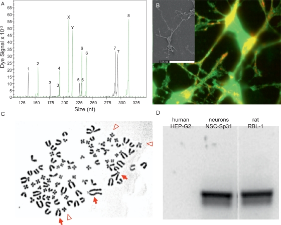
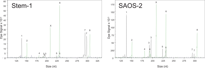
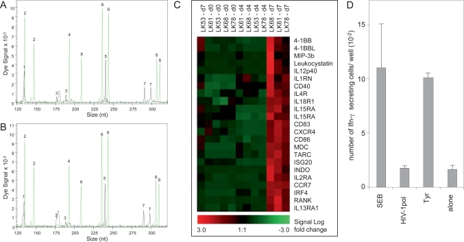
Cellular therapies that either use modifications of a patient's own cells or allogeneic cell lines are becoming in vogue. Besides the technical issues of optimal isolation, cultivation and modification, quality control of the generated cellular products are increasingly being considered to be more important. This is not only relevant for the cell's therapeutic application but also for cell science in general. Recent changes in editorial policies of respected journals, which now require proof of authenticity when cell lines are used, demonstrate that the subject of the present paper is not a virtual problem at all. In this article we provide 2 examples of contaminated cell lines followed by a review of the recent developments used to verify cell lines, stem cells and modifications of autologous cells. With relative simple techniques one can now prove the authenticity and the quality of the cellular material of interest and therefore improve the scientific basis for the development of cells for therapeutic applications. The future of advanced cellular therapies will require production and characterization of cells under GMP and GLP conditions, which include proof of identity, safety and functionality and absence of contamination.
Zelltherapien, die auf patienteneigenen Zellen oder allogenen Zellen basieren, werden zunehmend attraktiv und realisierbar. Neben technischen Fragen nach der optimalen Isolation und geeigneten Kultivierungsund Modifikationsverfahren tritt dabei auch die Bedeutung der Qualitätskontrolle für das Zellprodukt zunehmend ins Bewusstsein. Diese Entwicklung ist nicht nur bei Zellen für die therapeutische Anwendung wichtig, sondern auch für die Zellbiologie als Wissenschaft im Allgemeinen. Dass dies nicht nur ein virtuelles Problem ist, wird schon dadurch deutlich, dass angesehene Zeitschriften ihre Veröffentlichungspolitik geändert haben und nun einen Nachweis der Authentizität der verwendeten Zellen als Voraussetzung für die Veröffentlichung fordern. In diesem Beitrag stellen wir zwei Beispiele für die Kontamination von Zelllinien vor, gefolgt von einem Überblick über neuere Entwicklungen für die Verifikation von Zelllinien, Stammzellen und Zellmodifikationen. Mit relativ einfachen Techniken ist es heute möglich, die Authentizität der Zellen nachzuweisen, die Qualität des Zellmaterials zu beschreiben und damit die wissenschaftliche Basis für die Entwicklung von Zellen für die Therapie zu verbessern. Die Zukunft der sogenannten «Advanced Cellular Therapies» wird die Einhaltung von GMP- und GLP-Bedingungen erfordern. Dies schließt den Nachweis der Identität, Sicherheit und Funktionalität und der Freiheit von Kontamination für das Zellprodukt ein.
Figures
References
-
- Cheng L, Xiao L, Zeng F, Zhang YA. Stem cells shine in Shanghai. Cell Stem Cell. 2008;2:34–37. - PubMed
-
- Hay RJ. Cell Quantitation and characterization. In: Atala A, Lanza RP, editors. Methods of Tissue Engineering. San Diego: Academic Press; 2002. pp. 55–84.
-
- EU Regulation (EC) No 1394/2007 of the European Parliament and of the Council of 13 November 2007 on Advanced Therapy Medicinal Products and Amending Directive 2001/83/EC and Regulation (EC) No 726/2004. Official Journal of the European Union. 2007:121–137.
-
- Stacey GN. Cell contamination leads to inaccurate data: we must take action now. Nature. 2000;403:356. - PubMed
-
- Stacey GN. Standardisation of cell lines. Dev Biol (Basel) 2002;111:259–272. - PubMed
LinkOut - more resources
Full Text Sources
