. 2010 Dec;62(12):3574-83.
doi: 10.1002/art.27720.

Regulation of complement by cartilage oligomeric matrix protein allows for a novel molecular diagnostic principle in rheumatoid arthritis

Affiliations

Regulation of complement by cartilage oligomeric matrix protein allows for a novel molecular diagnostic principle in rheumatoid arthritis

Kaisa E Happonen et al. Arthritis Rheum. 2010 Dec.

Abstract

Objective: Cartilage oligomeric matrix protein (COMP) is a structural component of cartilage, where it catalyzes collagen fibrillogenesis. Elevated amounts of COMP are found in serum during increased turnover of cartilage associated with active joint disease, such as rheumatoid arthritis (RA) and osteoarthritis (OA). This study was undertaken to investigate the ability of COMP to regulate complement, a capacity that has previously been shown for some other cartilage proteins.

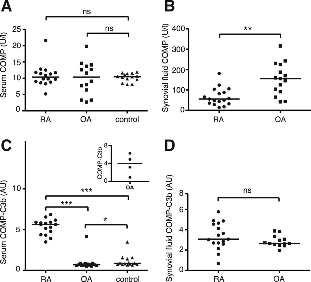
Methods: Regulation of complement by COMP was studied using functional in vitro assays. Inter-actions between complement proteins and COMP were investigated by direct binding assay and electron microscopy. Circulating COMP and COMP-C3b complexes in serum and synovial fluid from RA and OA patients and healthy controls were measured with a novel enzyme-linked immunosorbent assay.

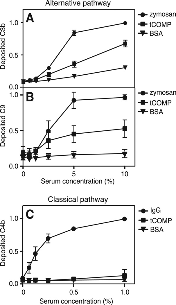
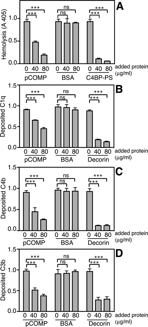
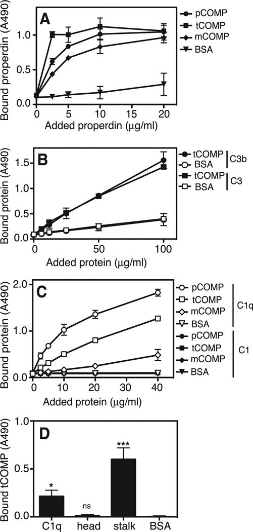
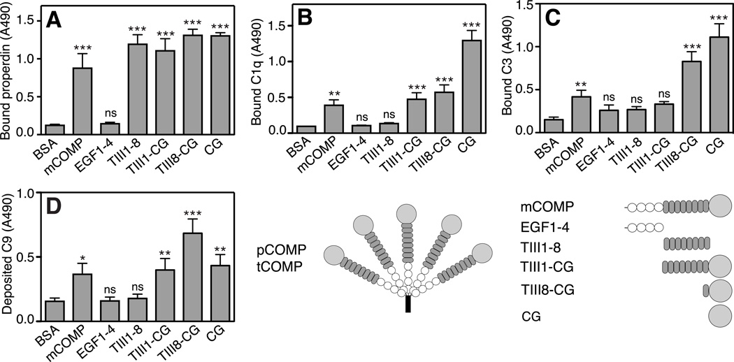
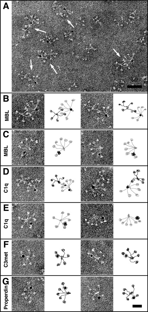
Results: We found in vivo evidence of complement activation by released COMP in the general circulation of patients with RA, but not patients with OA. COMP induced activation and deposition of C3b and C9 specifically via the alternative pathway of complement, which was attributable to direct interaction between COMP and properdin. Furthermore, COMP inhibited the classical and the lectin complement pathways due to direct interaction with the stalk region of C1q and mannose-binding lectin, respectively.

Conclusion: COMP is the first extracellular matrix protein for which an active role in inflammation has been demonstrated in vivo. It can activate one complement pathway at the same time as it has the potential to inhibit another. The net outcome of these interactions is most likely determined by the type of released COMP fragments, which may be disease specific.


Figures

FIGURE 1
FIGURE 1. COMP activates the alternative pathway of complement
tCOMP, zymosan and BSA were coated onto microtiter plates and increasing concentrations of NHS in Mg++EGTA were added. Activation of the alternative pathway was measured by detecting deposited C3b (A) and C9 (B). Activation of the classical and lectin pathways was measured by coating plates with tCOMP, aggregated human IgG or BSA and adding increasing concentrations of NHS in GVB++. Deposition of C4b was measured as an indication of complement activation (C). The graphs show the mean and standard deviation (SD) of three separate experiments. The data in the panels were normalized by setting the highest obtained absorbance of each plate to 1.
FIGURE 2
FIGURE 2. COMP inhibits the classical pathway of complement
The ability of pCOMP to inhibit erythrocyte lysis when pre-incubated with NHS was measured in a hemolytic assay. As the positive control, the main inhibitor of the classical pathway, C4BP-PS, was used (A). Plates were coated with aggregated human IgG and NHS pre-incubated with increas- ing concentrations of pCOMP was added. BSA and decorin were added to NHS as negative and positive controls. The panels show deposition of C1q (B), C4b (C) and C3b (D). The data are given as the mean and SD of three separate experiments and the data in panels B–D were normalized by setting the highest obtained absorbance of each plate to 1. Statistical significance of differences was calculated with a two-way ANOVA. ns, not significant, *, p < 0.05, **, p < 0.01, ***, p < 0.001.
FIGURE 3
FIGURE 3. COMP binds to properdin, C3 and C1q
tCOMP, pCOMP and mCOMP were coated onto microtiter plates and incubated with fluid phase properdin at increasing concentrations (A). To compare binding of C3 and the C3b activation product to tCOMP, tCOMP was immobilized and increasing concentrations of C3 and C3b were added to the plate. BSA was immobilized as a negative control (B). In order to compare binding of COMP to C1 and C1q, tCOMP, pCOMP and mCOMP were coated onto microtiter plates and fluid phase C1q or C1 at increasing concentrations was allowed to attach (C). To examine the binding region for COMP on C1q, isolated head and stalk fragments of C1q or intact C1q were immobilized and incubated with 20 µg/ml tCOMP (D). The data are given as the mean and SD of three separate experiments. Statistical significance was calculated using a one-way ANOVA in panel D. ns, not significant, *, p < 0.05, **, p < 0.01, ***, p < 0.001.
FIGURE 4
FIGURE 4. Binding of complement factors to COMP constructs
COMP-constructs were immobilized onto a microtiter plate from solutions of equimolar concentrations and incubated with fluid phase properdin (A), C1q (B) and C3 (C). C9-deposition through the alternative pathway on COMP constructs was measured by immobilizing COMP-constructs and adding NHS diluted in Mg++-EGTA (E). The data are given as the mean and SD of three separate experiments. Statistical significance was calculated with a one-way ANOVA by comparing binding or deposition of complement components onto COMP constructs compared to binding or deposition onto the negative control, BSA. ns, not significant, *, p < 0.05, **, p < 0.01, ***, p < 0.001.
FIGURE 5
FIGURE 5. Electron microscopy of COMP and complement proteins
Complexes between pCOMP and gold-labeled complement proteins or gold-labeled mCOMP and complement proteins were visualized by negative staining. A representative field of complexes between pCOMP and gold-labeled MBL is shown in (A). pCOMP in complex with MBL at a higher magnification (B). Selected particles of gold-labeled mCOMP bound to the arms of MBL (C). pCOMP in complex with gold-labeled C1q (D). Gold-labeled mCOMP in complex with C1q (E). Gold-labeled C3met (F) and properdin (G) bound to pCOMP. The schematic illustrations next to each image show COMP in black and complement proteins in grey. The scale bar represents 100 nm in (A) and 50 nm in (B–G).
FIGURE 6
FIGURE 6. COMP and COMP-C3b concentrations in RA and OA patients
The amount of released COMP in serum of RA patients (n=16) and OA patients (n=14) was measured using a commercial COMP® ELISA (A). COMP-concentrations were also measured in synovial fluid of RA patients (n=17) and OA patients (n=15) (B). Circulating complexes of COMP and C3b were measured using a sandwich ELISA in the serum of 16 RA patients, 14 OA patients and 14 healthy controls (C). The inset shows the levels of COMP-C3b in four OA patients with additional rheumatologi- cal disorders. COMP-C3b concentrations in synovial fluid were measured in 17 RA patients and 13 OA patients (D). The horizontal line in each panel represents the median. Statistical differences between the groups in panels A and C were determined using a Kruskal-Wallis test whereas a Mann-Whitney test was used in panels B and D. AU, arbitrary units; ns, not significant; *, p < 0.05, **, p < 0.01, ***, p < 0.001.

Similar articles

Cited by

References

    1. Hedbom E, Antonsson P, Hjerpe A, Aeschlimann D, Paulsson M, Rosa-Pimentel E, et al. Cartilage matrix proteins. An acidic oligomeric protein (COMP) detected only in cartilage. J Biol Chem. 1992;267(9):6132–6136. - PubMed
    1. Smith RK, Zunino L, Webbon PM, Heinegård D. The distribution of cartilage oligomeric matrix protein (COMP) in tendon and its variation with tendon site, age and load. Matrix Biol. 1997;16(5):255–271. - PubMed
    1. DiCesare P, Hauser N, Lehman D, Pasumarti S, Paulsson M. Cartilage oligomeric matrix protein (COMP) is an abundant component of tendon. FEBS Lett. 1994;354(2):237–240. - PubMed
    1. Dodge GR, Hawkins D, Boesler E, Sakai L, Jimenez SA. Production of cartilage oligomeric matrix protein (COMP) by cultured human dermal and synovial fibroblasts. Osteoarthritis Cartilage. 1998;6(6):435–440. - PubMed
    1. Saxne T, Heinegård D. Cartilage oligomeric matrix protein: a novel marker of cartilage turnover detectable in synovial fluid and blood. Br J Rheumatol. 1992;31(9):583–591. - PubMed

Publication types

MeSH terms