Review
. 2010 Oct 1;518(19):3883-906.
doi: 10.1002/cne.22435.

Central respiratory chemoreception

Affiliations
Review

Central respiratory chemoreception

Patrice G Guyenet et al. J Comp Neurol. .

Abstract

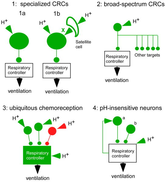
By definition central respiratory chemoreceptors (CRCs) are cells that are sensitive to changes in brain PCO(2) or pH and contribute to the stimulation of breathing elicited by hypercapnia or metabolic acidosis. CO(2) most likely works by lowering pH. The pertinent proton receptors have not been identified and may be ion channels. CRCs are probably neurons but may also include acid-sensitive glia and vascular cells that communicate with neurons via paracrine mechanisms. Retrotrapezoid nucleus (RTN) neurons are the most completely characterized CRCs. Their high sensitivity to CO(2) in vivo presumably relies on their intrinsic acid sensitivity, excitatory inputs from the carotid bodies and brain regions such as raphe and hypothalamus, and facilitating influences from neighboring astrocytes. RTN neurons are necessary for the respiratory network to respond to CO(2) during the perinatal period and under anesthesia. In conscious adults, RTN neurons contribute to an unknown degree to the pH-dependent regulation of breathing rate, inspiratory, and expiratory activity. The abnormal prenatal development of RTN neurons probably contributes to the congenital central hypoventilation syndrome. Other CRCs presumably exist, but the supportive evidence is less complete. The proposed locations of these CRCs are the medullary raphe, the nucleus tractus solitarius, the ventrolateral medulla, the fastigial nucleus, and the hypothalamus. Several wake-promoting systems (serotonergic and catecholaminergic neurons, orexinergic neurons) are also putative CRCs. Their contribution to central respiratory chemoreception may be behavior dependent or vary according to the state of vigilance.


Figures

Figure 1
Figure 1. what is a central respiratory chemoreceptor (CRC)?
The concept of central respiratory chemoreceptors is still being refined as more pertinent experimental evidence accumulates. Schemes 1-3 represent various CRC concepts. Scheme 4 represents neurons that should not be considered as CRCs. Specialized CRCs (option 1) could be defined as neurons that detect the concentration of protons in the surrounding brain parenchyma and drive the respiratory rhythm and pattern generating network selectively and in a graded manner according to the level of acidity. Option 1a assumes that these specialized neurons express the proton receptors. Option 1b assumes that the proton receptors reside both on these specialized neurons and on satellite cells (e.g. glia) that communicate with the neurons via a messenger X (possibly ATP). In this second case, the satellite cells would also qualify as CRCs. Broad-spectrum CRCs (option 2) could be defined as proton-sensitive neurons that modulate the activity of numerous targets besides the respiratory controller. Subsets of serotonergic neurons, the locus coeruleus and orexin neurons may be in this category. Ubiquitous chemoreception (option 3) refers to the possibility that central respiratory chemoreception could be an emergent property caused by the summation of small effects of pH almost everywhere in the respiratory network. Option 4 illustrates hypothetical examples of neurons that regulate the respiratory controller and its reactivity to protons. These neurons do not qualify as CRCs because they are not directly affected by the extracellular proton concentration.
Figure 2
Figure 2. the retrotrapezoid nucleus (RTN)
A. The RTN contains a neurochemically well-defined group of glutamatergic neurons, the ccRTN neurons (Takakura et al., 2008; Lazarenko et al., 2009) so named to distinguish them from the overlapping but phenotypically heterogeneous collection of cells called the parafacial respiratory group (Guyenet and Mulkey, 2010). The axonal projections of the ccRTN neurons in the rat are represented schematically based on the anterograde transport of the membrane bound fusion protein channelrhodopsin2-mCherry (for primary data see (Abbott et al., 2009). The scale (1 mm) is an approximation based on the rostrocaudal extent of the facial motor nucleus in the adult rat. B. transverse histological section along the plane identified by the dotted line in panel A (after (Stornetta et al., 2006)). The ccRTN neurons express the transcription factor Phox2b (nuclei in green) and lack tyrosine hydroxylase (TH, in red). The TH-ir neurons located at this level are the C1 neurons, which regulate the heart and vasomotor tone. The location of the facial motor neurons (7) is revealed by the presence of fluorogold (in blue). The scale bar is 100 microns. The ventral medullary surface is recognizable by a green edge artifact. C. CO2-sensitivity of the ccRTN neurons in an anesthetized rat in vivo. C1: experimental design for unit recording in anesthetized rats. The ccRTN neurons are located under the facial motor nucleus the lower boundary of which is identified by antidromic field potentials elicited by stimulation of the facial nerve. C2: single ccRTN neuron recorded alongside end-expiratory CO2 (top trace), arterial blood pressure (AP) and the mass activity of the phrenic nerve (iPND; a reporter of the level of activity of the respiratory controller). ccRTN neurons are robustly activated by adding CO2 to the breathing mixture. The expanded time-scale insert shows that the neuron has only a mild propensity to discharge in synchrony with the central respiratory controller. C3 is an example of one such neuron labeled in vivo with biotinamide following its physiological characterization. Note the profusion of dendrites at the ventral medullary surface (VMS) (after (Mulkey et al., 2004). D: selective stimulation of the ccRTN neurons activates breathing in vivo. D1, experimental design. A thin optical fiber is introduced in the vicinity of a group of ccRTN neurons that were selectively transfected with the channelrhodopsin2-mCherry (ChR2) fusion protein. ccRTN neurons are recorded while blue laser light is applied to activate ChR2-expressing neurons. The rat is anesthetized with isoflurane. D2, transverse section showing several superficial Phox2b-expressing ccRTN neurons that were transfected with ChR2 (m-Cherry exhibits a natural red fluorescence). The transgene was expressed almost exclusively by Phox2b-containing neurons (Phox2b immunoreactivity appears green in untransfected neurons, green arrow, and yellow in transfected cells, white arrows). The transfected neurons line the ventral medullary surface. Note that the overlying facial motor neurons (7) did not express the transgene (scale bar: 0.1 mm). D3, activation of ChR2-transfected ccRTN neurons (one of them is being simultaneously recorded) activates breathing. In this excerpt, the end-expiratory CO2 was below the apneic threshold therefore both the RTN unit and the phrenic nerve were silent prior to shining the laser light (after (Abbott et al., 2009)). During light application, the RTN unit was entrained 1 to 1 to the laser pulses (bottom two traces). E: pH sensitivity of ccRTN neurons in brain slices. E1, ccRTN neurons in a Phox2b-eGFP transgenic mouse (the B/G mouse). In this strain, GFP identifies selectively the ccRTN neurons (scale bar: 50 microns). E2, thick section (∼300 μm) through the RTN of the B/G mouse. Two ccRTN neurons were recorded in vitro and were filled with biotinamide (scale bar: 100 microns). E3, typical response of a ccRTN neuron to acidification in vitro (B/G mouse, temperature: 22°C). The cell was silent at pH 7.5. It was activated by acidification and by substance P (SP; 100 nM), the latter due to the presence of neurokinin1 receptors on the cells. Panels E1-3 after (Lazarenko et al., 2009). F1, ccRTN neurons fit the concept of specialized chemoreceptors. The molecular and cellular mechanism by which they detect surrounding protons (1a or 1b) is uncertain however. F2, a slightly more elaborate concept of how ccRTN neurons might regulate breathing. ccRTN neurons drive the breathing rate, the amplitude of inspiration and, probably active expiration. ccRTN neurons regulate breathing according to the level of acidity that surrounds the neurons but also according to the strength of the input that they receive from the carotid bodies, the raphe and the hypothalamus, including from orexinergic neurons. Distinct subsets of ccRTN neurons may drive the various respiratory motor outflows (inspiratory vs. expiratory muscles).
Figure 3
Figure 3. serotonergic neurons and central respiratory chemosensitivity
A, ventral view of the rat brainstem showing the location of the serotonergic neurons that reside close to the ventral medullary surface (tryptophan-hydroxylase, TpOHase, immunoreactivity in green; calibration bar: 1.5 mm). The blood vessels are red because they have been filled with a resin. The superficial serotonergic neurons are close to blood vessels and reside in regions of the ventral medullary surface assumed to contain CRCs because experimental acidification of these regions increases breathing. We added the white ovals that outline the retrotrapezoid nucleus. B, typical serotonin neuron in culture that responds briskly to extracellular acidification. A and B reprinted with slight modification from Respiration Physiology and Neurobiology, Volume 168, Corcoran et al., Medullary serotonin neurons and central CO2 chemoreception, pp. 49-58, Copyright (2009), with permission from Elsevier. C. the lateral B3 group of serotonergic neurons is insensitive to hypercapnia in anesthetized rats. C1, example of a serotonergic neuron (tryptophan-hydroxylase-immunoreactive) filled with biotinamide after electrophysiological study in vivo (calibration bar: 20 microns). C2, location of 24 biochemically identified serotonergic neurons found unresponsive to hypercapnia in vivo (red dots). These neurons were located in the lateral band of serotonergic neurons shown in panel A at the level of the RTN (pyr, pyramidal tract; RPa, raphe pallidus; TpOHase, tryptophan hydroxylase). C3 example of a single identified lateral B3 group serotonergic neuron. The neuron was unaffected by increasing end-expiratory CO2 by 5% from just below the apneic threshold. The activity of the respiratory controller, monitored at the level of the phrenic nerve (iPND) was robustly activated by CO2 as expected. C1-3 from (Mulkey et al., 2004). D, raphe obscurus serotonergic neurons driven by the respiratory controller. These recordings were obtained in a coronal slice of neonate medulla oblongata that generates a respiratory-like activity (the “breathing slice”) (Smith et al., 1991). In this slice, the activity of the residual respiratory network was monitored by the mass discharge of the hypoglossal motor neurons (integral XII). In the lower two traces the membrane potential of the serotonergic cell was deliberately hyperpolarized to reveal the excitatory drive potential synchronized with the inspiratory phase. Reproduced with permission from the Journal of Neuroscience from (Ptak et al., 2009). E, putative raphe obscurus serotonergic neurons recorded in a conscious cat. Most cells (21/27) did not respond to hypercapnia. Small subsets of putative serotonergic neurons, one of which is illustrated here, were activated by CO2 but only while the cats were awake. Reproduced with permission from the Journal of Neuroscience from (Veasey et al., 1995). F, the release of serotonin contributes to the activity of the respiratory controller in vitro. The figure shows the mass activity of the preBötzinger region of the ventral respiratory column in the “breathing slice” (ipreBot, an indication of the inspiratory phase of the breathing cycle; top trace) and an inspiratory neuron that was recorded intracellularly (lower trace). Superfusion of the slice with a serotonin receptor 2A antagonist slowed the fictive breathing rate and the amplitude of the respiratory bursts indicating that the ongoing release of serotonin was contributing to the activity of the network under these in vitro conditions. Reproduced with permission from the Journal of Neuroscience from (Pena and Ramirez, 2002). G, putative role of serotonergic neurons in central chemoreception. Left: a small subset of serotonergic neurons, the location of which needs to be clarified, may be central respiratory chemoreceptors, i.e. may be acid-sensitive in vivo and may activate the respiratory controller among other targets. Right, most serotonergic neurons recorded so far in adult mammals in vivo were not CO2-responsive. A few serotonergic neurons were CO2–activated, either because of an intrinsic acid-sensitivity or because they receive excitatory inputs from the respiratory controller. The serotonergic system at large activates the breathing network and regulates its response to CO2. RTN neurons receive an excitatory input from serotonergic cells (Mulkey et al., 2007a). In theory, this input could originate from pH-sensitive (left) or pH-insensitive serotonergic cells (right).

Similar articles

Cited by

References

    1. Abbott SBG, Stornetta RL, Fortuna MG, Depuy SD, West GH, Harris TE, Guyenet PG. Photostimulation of retrotrapezoid nucleus Phox2b-expressing neurons in vivo produces long-lasting activation of breathing in rats. J Neurosci. 2009;29:5806–5819. - PMC - PubMed
    1. Abdala AP, Rybak IA, Smith JC, Paton JF. Abdominal expiratory activity in the rat brainstem-spinal cord in situ: patterns, origins, and implications for respiratory rhythm generation. J Physiol. 2009 doi: 10.113/jphysiol.2008.167502. - DOI - PMC - PubMed
    1. Ahmad HR, Loeschcke HH. Transient and steady state responses of pulmonary ventilation to the medullary extracellular pH after approximately rectangular changes in alveolar PCO2. Pflugers Arch. 1982;395:285–292. - PubMed
    1. Ainslie PN, Duffin J. Integration of cerebrovascular CO2 reactivity and chemoreflex control of breathing: mechanisms of regulation, measurement, and interpretation. Am J Physiol Regul Integr Comp Physiol. 2009;296:R1473–R1495. - PubMed
    1. Aller MI, Wisden W. Changes in expression of some two-pore domain potassium channel genes (KCNK) in selected brain regions of developing mice. Neurosci. 2008;151:1154–1172. - PubMed

Publication types