

Dorsal epidural intervertebral disk herniation with atypical radiographic findings: case report and literature review
- PMID: 20737802
- PMCID: PMC2920122
- DOI: 10.1080/10790268.2010.11689706
Dorsal epidural intervertebral disk herniation with atypical radiographic findings: case report and literature review
Abstract
Background/objective: Intervertebral disk herniation is relatively common. Migration usually occurs in the ventral epidural space; rarely, disks migrate to the dorsal epidural space due to the natural anatomical barriers of the thecal sac.
Design: Case report.
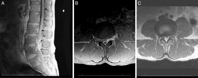
Findings: A 49-year-old man presented with 1 week of severe back pain with bilateral radiculopathy to the lateral aspect of his lower extremities and weakness of the ankle dorsiflexors and toe extensors. Lumbar spine magnetic resonance imaging with gadolinium revealed a peripheral enhancing dorsal epidural lesion with severe compression of the thecal sac. Initial differential diagnosis included spontaneous hematoma, synovial cyst, and epidural abscess. Posterior lumbar decompression was performed; intraoperatively, the lesion was identified as a large herniated disk fragment.
Conclusions: Dorsal migration of a herniated intervertebral disk is rare and may be difficult to definitively diagnose preoperatively. Dorsal disk migration may present in a variety of clinical scenarios and, as in this case, may mimic other epidural lesions on magnetic resonance imaging.
Figures
References
-
- Bonaroti EA, Welch WC. Posterior epidural migration of an extruded lumbar disk fragment causing cauda equina syndrome: clinical and magnetic resonance imaging evaluation. Spine. 1998;23(3):378–381. - PubMed
-
- Kuzeyli K, Cakir E, Usul H, et al. Posterior epidural migration of lumbar disk fragments: report of three cases. Spine. 2003;28(3):E64–E67. - PubMed
-
- Schellinger D, Manz HJ, Vidic B, et al. Disk fragment migration. Radiology. 1990;175(3):831–836. - PubMed
-
- Ebeling U, Reulen HJ. Are there typical localisations of lumbar disk herniations? A prospective study. Acta Neurochir. 1992;117(3–4):143–148. - PubMed
-
- Sekerci Z, Ildan F, Yuksel M, Gul B, Kilic C. Cauda equina compression due to posterior epidural migration of extruded lumbar disk. Neurosurg Rev. 1992;15(4):311–313. - PubMed
Publication types
MeSH terms
Substances
LinkOut - more resources
Full Text Sources
Medical