. 2011 Jan;132(1):27-38.
doi: 10.1111/j.1365-2567.2010.03334.x. Epub 2010 Aug 25.

Human cytomegalovirus-specific CD8(+) T-cell expansions contain long-lived cells that retain functional capacity in both young and elderly subjects

Affiliations

Human cytomegalovirus-specific CD8(+) T-cell expansions contain long-lived cells that retain functional capacity in both young and elderly subjects

Diana L Wallace et al. Immunology. 2011 Jan.

Abstract

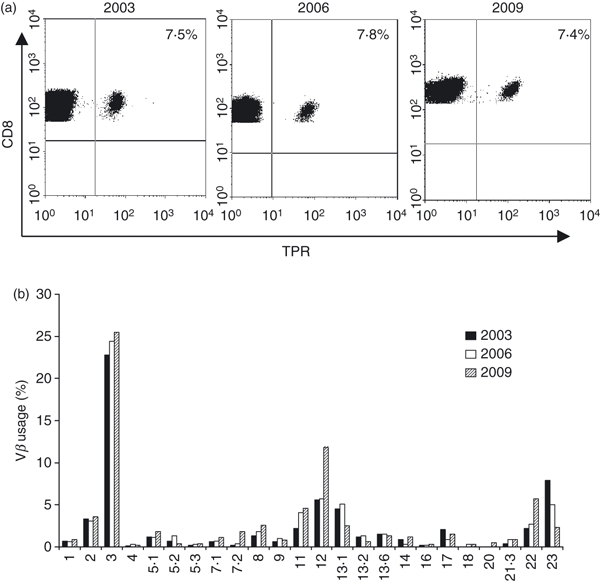
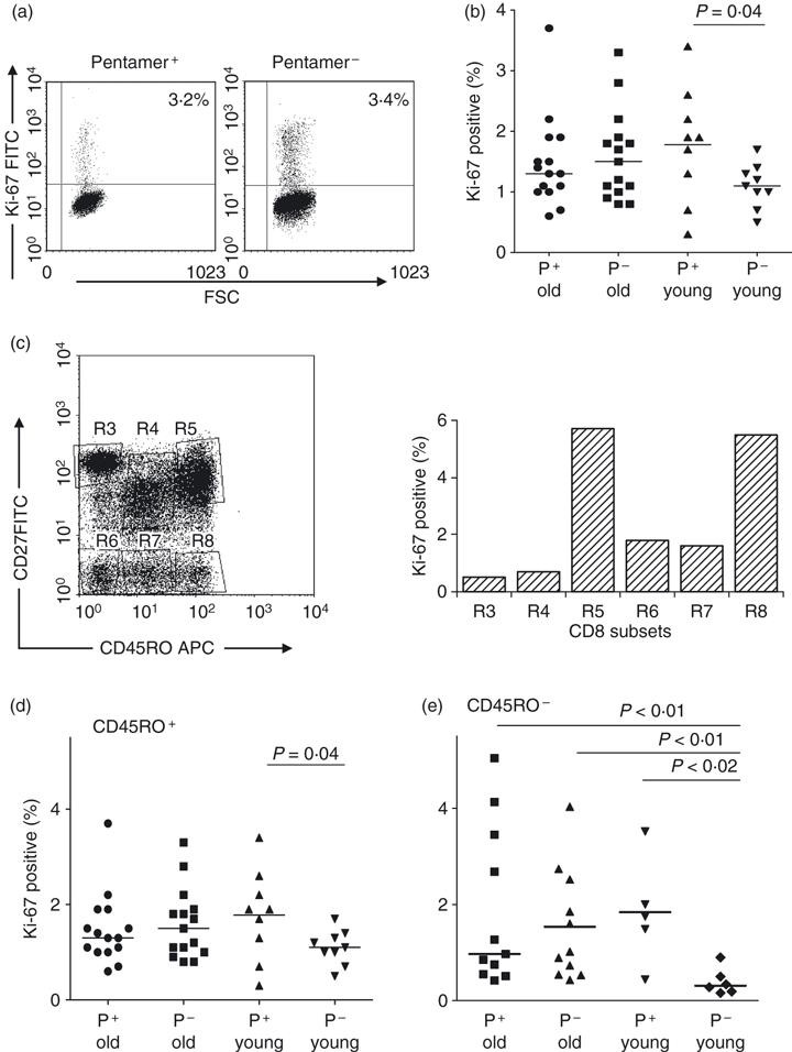
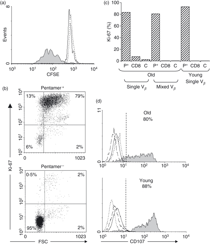
The immune response to human cytomegalovirus (HCMV) infection is characterized by the accumulation of HCMV-specific CD8(+) T cells, particularly in the elderly; such expansions may impair immune responses to other pathogens. We investigated mechanisms underlying HCMV-specific expansions in 12 young and 21 old healthy subjects (although not all analyses were performed on all subjects). Phenotypically, HCMV-pentamer(+) CD8(+) T cells were characterized by marked Vβ restriction, advanced differentiation (being predominantly CD27(-) CD28(-) ), and variable CD45RO/RA expression. Although more common and larger in older subjects, expansions had similar phenotypic characteristics in the young. In one old subject, repeated studies demonstrated stability in size and Vβ distribution of pentamer(+) populations over 6 years. We tested whether HCMV-specific CD8(+) T-cell expansions arose from accelerated proliferation or extended lifespan by in vivo labelling with deuterated glucose and ex vivo Ki-67 expression. Uptake of deuterated glucose was lower in pentamer(+) cells than in pentamer(-) CD8(+) CD45RO(+) or CD8(+) CD45RA(+) cells in three old subjects, consistent with reduced proliferation and extended lifespan. Similarly Ki-67 labelling showed no evidence for increased proliferation in HCMV-specific CD8(+) expansions in older subjects, although pentamer(-) CD45RA(+) cells from young donors expressed very little Ki-67. We investigated Bcl-2 and CD95 as possible anti-apoptotic mediators, but neither was associated with pentamer-positivity. To investigate whether expansion represents a compensatory response to impaired functionality, we performed two tests of functionality, peptide-stimulated proliferation and CD107 expression; both were intact in pentamer(+) cells. Our data suggest that HCMV-specific CD8(+) expansions in older subjects accumulate by extended lifespan, rather than accelerated proliferation.


Figures

Figure 1
Figure 1
CD8+ human cytomegalovirus (HCMV) -specific T-cell receptor (TCR) Vβ expansions in young and old. Peripheral blood mononuclear cells from HCMV-seropositive donors were stained with MHC class I pentamers to determine the percentage of CD8+ HCMV-specific T cells. (a) Representative flow cytometric profiles from young and old donors showing the percentage of total CD8+ cells specific for the HCMV peptides NLV (donors O11 and Y5) or TPR (donors O2 and Y1). (b) Percentage of CD8+ HCMV-specific cells identified for all donors included in the study. The horizontal line represents the median value for each group (young and old for each peptide); comparisons are by Mann–Whitney U-test. (c) TCR Vβ usage of total CD8+ (open bars) and NLV-specific (solid bars) T cells in a representative old donor (O11); cells were co-stained with pentamer and 24 different anti-TCR Vβ antibodies (in order: Vβ 1, 2, 3, 4, 5.1, 5.2, 5.3, 7.1, 7.2, 8, 9, 11, 12, 13.1, 13.2, 13.6, 14, 16, 17, 18, 20, 21.3, 22, 23). (d) As described for (c) but for a representative young donor (Y5).
Figure 2
Figure 2
Long-term stability of human cytomegalovirus (HCMV) -specific expansions in an elderly subject. Peripheral blood mononuclear cells taken three times over a 6-year period from a single old HCMV+ B7-restricted donor (O3) were stained with (a) CD8 and MHC class I TPR-specific pentamer and (b) with CD8 and the same Vβ panel as detailed in Fig. 1.
Figure 3
Figure 3
In vivo proliferation and disappearance of CD8+ T cells. Fraction of labelled cells following deuterium-labelled glucose administration for 10 hr at time 0. Peak height indicates proliferation rate and rate of decline indicates disappearance rate. Pentamer+ cells (filled squares) were separated from CD45RO+ (open squares) and CD45RO (open circles) before analysis. Data are shown from two individual subjects, O13 (a) and O4 (b); expansions were predominantly CD45RO in O13 but CD45RO+ in O4.
Figure 4
Figure 4
Ex vivo proliferation measured by Ki-67 expression. Peripheral blood mononuclear cells from young and old human cytomegalovirus (HCMV) -positive donors were stained for CD8, specific pentamer and Ki-67 and gated on pentamer+ CD8+ T cells, control cells from an HCMV-negative old donor gated on CD8+ T cells were stained for Ki-67. (a) Representative dot plots comparing the pentamer+ CD8+ T-cell population with the CD8+ pentamer population from the same old donor (O2). (b) Comparison of Ki-67 expression on all CD8+ cells derived from old (n = 15) and young (n = 9) donors. P-value by Wilcoxon signed rank test. (c) Comparison of Ki-67 activity on subsets defined by CD27 × CD45RO staining. (d) Expression of Ki-67 on subsets of CD45RO+ cells stained with CD8, MHC class I HCMV-specific pentamer and CD45RO from panels of old (n = 15) and young (n = 9) donors. Each data point represents the value from one donor; horizontal bars are medians. P-value quoted is by pairwise comparison (Wilcoxon signed rank test); no other significant differences between groups by analysis of variance (anova; Kruskal–Wallace). (e) As for (d) but for CD45RO cells (n = 11 and six respectively). P-value by anova (Kruskal–Wallace) = 0.019. Comparisons between groups by Mann–Whitney U-test.
Figure 5
Figure 5
Altered expression of Bcl-2 and CD95 does not explain the extended lifespan of CD8+ human cytomegalovirus (HCMV) -specific T cells within T-cell receptor (TCR) Vβ expansions. Peripheral blood mononuclear cells from four young and four old subjects were stained with MHC class I pentamers to identify HCMV-specific CD8+ T cells and the apoptotic potential of this population was assessed by flow cytometry after staining for CD95 and Bcl-2. Representative flow cytometric profiles from one old (left panel) and one young (right panel) donor showing CD95 (upper row, donors O15 and Y5) and Bcl-2 (bottom row donors O14 and Y8) expression on HCMV pentamer+ cells. Plots are gated on total CD8+ T cells.
Figure 6
Figure 6
Proliferative responses to human cytomegalovirus (HCMV) peptides and functional capacity of HCMV-specific CD8+ T cells from old and young. (a) Peripheral blood mononuclear cells (PBMC) from an HCMV+ A2-restricted donor (donor O11) with an NLV+ CD8+ T cell expansion, consisting predominantly of Vβ16, were labelled with CFSE and incubated with NLV peptide for 6 days before analysis on a FACScalibur. Solid fill represents CD8+ NLV+ peptide-stimulated cells on day 6, solid line represents CD8+ NLV+ cells on day 0 and dashed line represents CD8+ NLV peptide-stimulated cells on day 6. (b) PBMC from an old donor with a pentamer+ population containing a small Vβ expansion (donor O14) were stimulated with specific HCMV peptide for 3 days and then stained for Ki-67 (dot plot). (c) Histogram of Ki-67 expression in stimulated cells from one old donor (O6) one with a single dominant Vβ expansion and one young donor (Y9) with a dominant Vβ expansion; P+, pentamer+ cells; CD8, pentamer CD8+ cells, C, isotype control. (d) The functional profile of HCMV-specific CD8+ T cells was assessed in response to peptide stimulation by measuring surface mobilization of CD107a by flow cytometry. PBMC from one old (donor O6) and one young HLA-B7+ donor (Y1) were stimulated for 3 days with HCMV-B7 restricted peptides then stained with CD107 monoclonal antibodies, incubated with monensin for 6 hr, then stained with TPR and CD8 mAb. Solid fill represents CD8+ TPR+ peptide-stimulated cells; solid line represents control CD8+ TPR peptide-stimulated cells; dotted line represents CD8+ TPR+ isotype; dashed line represents TPR+ CD8+ cells incubated in medium (no peptide) for 3 days. Data are representative of studies in two young and four elderly donors.

References

    1. Sinclair J, Sissons P. Latency and reactivation of human cytomegalovirus. J Gen Virol. 2006;87:1763–79. - PubMed
    1. Waller EC, Day E, Sissons JG, Wills MR. Dynamics of T cell memory in human cytomegalovirus infection. Med Microbiol Immunol. 2008;197:83–96. - PubMed
    1. Klenerman P, Hill A. T cells and viral persistence: lessons from diverse infections. Nat Immunol. 2005;6:873–9. - PubMed
    1. Pawelec G, Larbi A, Derhovanessian E. Senescence of the human immune system. J Comp Pathol. 2010;142S:S39–44. - PubMed
    1. Pawelec G, Akbar A, Caruso C, Solana R, Grubeck-Loebenstein B, Wikby A. Human immunosenescence: is it infectious? Immunol Rev. 2005;205:257–68. - PubMed

Publication types

MeSH terms

Substances