Review
. 2010 Oct;14(10):2381-92.
doi: 10.1111/j.1582-4934.2010.01164.x.

Clinical characteristics and pathophysiological mechanisms of focal and diffuse traumatic brain injury

Affiliations
Review

Clinical characteristics and pathophysiological mechanisms of focal and diffuse traumatic brain injury

Teuntje M J C Andriessen et al. J Cell Mol Med. 2010 Oct.

Abstract

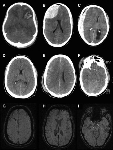
Traumatic brain injury (TBI) is a frequent and clinically highly heterogeneous neurological disorder with large socioeconomic consequences. TBI severity classification, based on the hospital admission Glasgow Coma Scale (GCS) score, ranges from mild (GCS 13-15) and moderate (GCS 9-12) to severe (GCS ≤ 8). The GCS reflects the risk of dying from TBI, which is low after mild (∼1%), intermediate after moderate (up to 15%) and high (up to 40%) after severe TBI. Intracranial damage can be focal, such as epidural and subdural haematomas and parenchymal contusions, or diffuse, for example traumatic axonal injury and diffuse cerebral oedema, although this distinction is somewhat arbitrary. Study of the cellular and molecular post-traumatic processes is essential for the understanding of TBI pathophysiology but even more to find therapeutic targets for the development of neuroprotective drugs to be eventually used in human beings. To date, studies in vitro and in vivo, mainly in animals but also in human beings, are unravelling the pathological TBI mechanisms at high pace. Nevertheless, TBI pathophysiology is all but completely elucidated. Neuroprotective treatment studies in human beings have been disappointing thus far and have not resulted in commonly accepted drugs. This review presents an overview on the clinical aspects and the pathophysiology of focal and diffuse TBI, and it highlights several acknowledged important events that occur on molecular and cellular level after TBI.


Figures

Fig 1
Fig 1
Focal and diffuse TBI. Examples of focal and diffuse TBI on CT (rows 1 and 2) and MRI (row 3). Focal injury: (A) Left frontal contusion with midline shift to the right and compression of the lateral ventricles. (B) Right frontal epidural haematoma with midline shift to the left and compression of the anterior part of the lateral ventricle. (C) Right frontotemporoparietal subdural haematoma with a midline shift to the left. Diffuse injury: (D) Punctate haemorrhage within the right posterior limb of the internal capsule, a sign of DAI; (E) Diffuse subarachnoid haemorrhage; (F) Diffuse swelling with bilateral compression of the basal cisterns. DAI on MRI: Susceptibility weighted images of one patient revealing punctate haemorrhages (hypo-intense foci) within (G) the right frontal hemisphere, (H) Splenium of the corpus callosum, and (I) mesencephalon, corresponding to grade 3 DAI.
Fig 3
Fig 3
Mechanical and pathophysiological mechanisms of TAI. Sudden lateral acceleration– deceleration of the head (A) is the main injury mechanism causing TAI. (B) and (C) are schematic representations of the brain during rest and movement, respectively. Deformation of the brain during sudden head movement causes shear, tensile and compressive strains within the brain tissue. Predilection sites of axonal injury are the grey and white matter interface, the corpus callosum and the brain stem (C). Sufficiently high strains, as occur, for example in motor-vehicle accidents, cause a cascade of pathological changes within the axon that may finally lead to axonal disconnection (D). It is suggested that two different processes of axonal injury exist [110]. The first is characterized by altered focal axolemma permeability (D1.I) whereby ionic homeostasis leads to local Ca2+ influx and mitochondrial swelling. Both local calcium dysregulation and release of cytochrome-c from damaged mitochondria result in activation of cysteine proteases and breakdown of essential axonal cytoskeleton products including loss of microtubules, neurofilament side-arm cleavage and neurofilament compaction impending normal axonal transport. In contrast to previous suggestions there is no termination of axonal transport or axonal swelling. Rather, it is suggested that there is a conversion of anterograde into retrograde axonal transport that prevents the axon from swelling. The second type of axonal injury (D1.II) is characterized by a combination of local axonal swelling and altered axonal transport but no overtly altered axolemma permeability. It is suggested that with this injury type there may be subtle alterations of membrane permeability triggering the activation of calcineurin. Calcineurin in turn alters the microtubular network, causing a disruption in axonal transport, with accumulation of organelles and swelling. After axonal disconnection, which may occur after both injury types, the axon undergoes a process of Wallerian degeneration (D2) consisting of a breakdown of the myelin sheath and the axon cylinder. The target site has now lost its input from the disconnected axon (D3) and may undergo synaptic reorganization, for example through axonal sprouting of neighbouring intact fibres. This process of synaptic reorganization may by adaptive or maladaptive.
Fig 2
Fig 2
Simplified pathophysiological molecular and cellular processes after focal TBI. The figure accompanies the transcript given in the main text. In short, increases in extracellular glutamate result in a supraphysiological Ca2+ influx that, subsequently, initiates several in parallel operating intracellular cascades (four are shown). Left upper corner: Increased activity of the calcium-dependent enzymes nNOS and eNOS enhances nitric oxide production leading to lipid peroxidation and eventually cellular necrosis. Left middle part: Calpain (a cystein protease) activity is also augmented due to an intracellular calcium increase, ultimately resulting in cellular necrosis pathways. Lysosomal membrane rupture and cathepsin release play an important role in this process. Right upper corner: Increases in intracellular calcium causing mitochondrial calcium overload result in increased mitochondrial membrane permeability. As a result ROS causing oxidative stress and the protein cytochrome-c are released into the cytoplasm. Cytochrome-c binds to the apoptosis activating protein-1 (apaf-1) activating the apoptotis inducing caspase pathway. Glu: Glutamate; [Glu]e: extracellular glutamate concentration; NMDA: N-methyl-D-aspartate aspartic acid; E.R.: endoplasmatic reticulum; [Ca2+]i: intracellular Ca2+ concentration; nNOS: neuronal NOS; eNOS: endothelial NOS; MPT: membrane permeability transition; ATP: adenosine triphosphate; ADP: adenosine diphosphate; ROS: reactive oxygen species; apaf-1: apoptosis activating protein-1;. O2: oxygen radical.

Similar articles

Cited by

References

    1. Tagliaferri F, Compagnone C, Korsic M, et al. A systematic review of brain injury epidemiology in Europe. Acta Neurochir. 2006;148:255–68. - PubMed
    1. Rutland-Brown W, Langlois JA, Thomas KE, et al. Incidence of traumatic brain injury in the United States, 2003. J Head Trauma Rehabil. 2006;21:544–8. - PubMed
    1. Murray GD, Teasdale GM, Braakman R, et al. The European Brain Injury Consortium survey of head injuries. Acta Neurochir. 1999;141:223–36. - PubMed
    1. Cremer OL, Moons KG, Van Dijk GW, et al. Prognosis following severe head injury: development and validation of a model for prediction of death, disability, and functional recovery. J Trauma. 2006;61:1484–91. - PubMed
    1. Myburgh JA, Cooper DJ, Finfer SR, et al. Epidemiology and 12-month outcomes from traumatic brain injury in australia and new zealand. J Trauma. 2008;64:854–62. - PubMed