

The liposoluble proteome of Mycoplasma agalactiae: an insight into the minimal protein complement of a bacterial membrane
- PMID: 20738845
- PMCID: PMC2941501
- DOI: 10.1186/1471-2180-10-225
The liposoluble proteome of Mycoplasma agalactiae: an insight into the minimal protein complement of a bacterial membrane
Abstract
Background: Mycoplasmas are the simplest bacteria capable of autonomous replication. Their evolution proceeded from gram-positive bacteria, with the loss of many biosynthetic pathways and of the cell wall. In this work, the liposoluble protein complement of Mycoplasma agalactiae, a minimal bacterial pathogen causing mastitis, polyarthritis, keratoconjunctivitis, and abortion in small ruminants, was subjected to systematic characterization in order to gain insights into its membrane proteome composition.
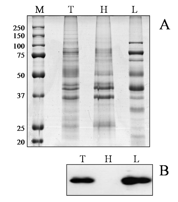
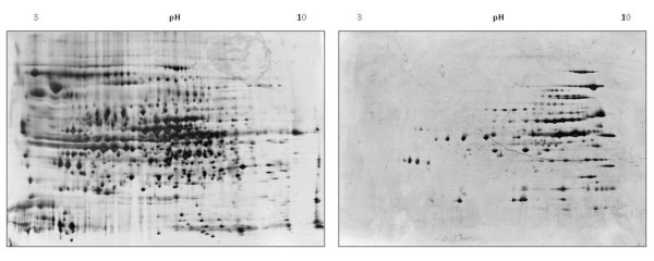
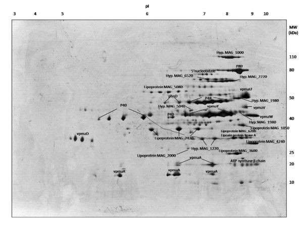
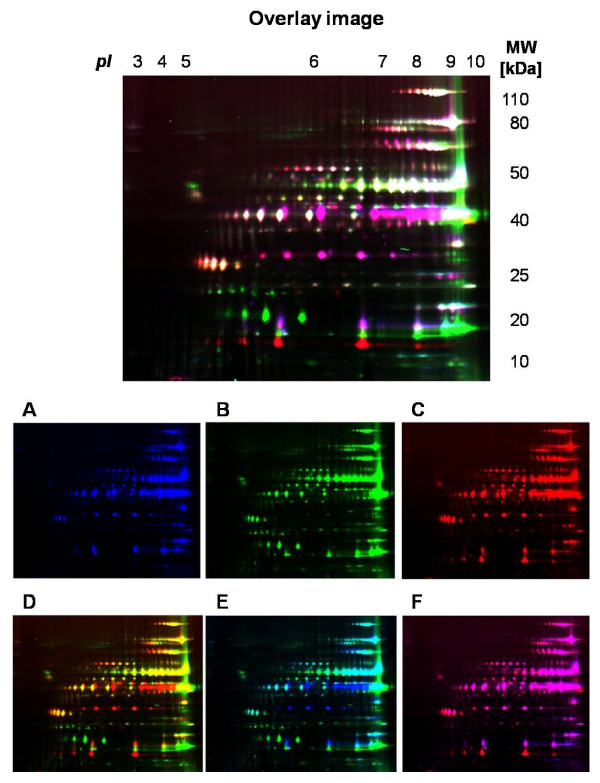
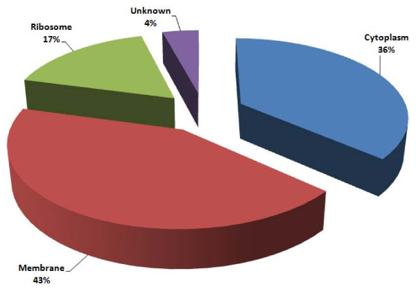
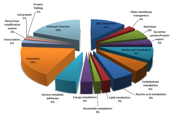
Results: The selective enrichment for M. agalactiae PG2T liposoluble proteins was accomplished by means of Triton X-114 fractionation. Liposoluble proteins were subjected to 2-D PAGE-MS, leading to the identification of 40 unique proteins and to the generation of a reference 2D map of the M. agalactiae liposoluble proteome. Liposoluble proteins from the type strain PG2 and two field isolates were then compared by means of 2D DIGE, revealing reproducible differences in protein expression among isolates. An in-depth analysis was then performed by GeLC-MS/MS in order to achieve a higher coverage of the liposoluble proteome. Using this approach, a total of 194 unique proteins were identified, corresponding to 26% of all M. agalactiae PG2T genes. A gene ontology analysis and classification for localization and function was also carried out on all protein identifications. Interestingly, the 11.5% of expressed membrane proteins derived from putative horizontal gene transfer events.
Conclusions: This study led to the in-depth systematic characterization of the M. agalactiae liposoluble protein component, providing useful insights into its membrane organization.
Figures
References
-
- Rottem S. Interaction of mycoplasmas with host cells. Physiol Rev. 2003;83:417–432. - PubMed
-
- Kühner S, van Noort V, Betts MJ, Leo-Macias A, Batisse C, Rode M, Yamada T, Maier T, Bader S, Beltran-Alvarez P, Castaño-Diez D, Chen WH, Devos D, Güell M, Norambuena T, Racke I, Rybin V, Schmidt A, Yus E, Aebersold R, Herrmann R, Böttcher B, Frangakis AS, Russell RB, Serrano L, Bork P, Gavin AC. Proteome organization in a genome-reduced bacterium. Science. 2009;27:1235–1240. doi: 10.1126/science.1176343. - DOI - PubMed
-
- Lambert M. Contagious agalactia of sheep and goats. Rev Sci Tech OIE. 1987;6:699–711. Mycoplasmoses of ruminants. - PubMed
