. 2010 Aug 25:11:63.
doi: 10.1186/1471-2199-11-63.

Functional and cellular characterization of human Retinoic Acid Induced 1 (RAI1) mutations associated with Smith-Magenis Syndrome

Affiliations

Functional and cellular characterization of human Retinoic Acid Induced 1 (RAI1) mutations associated with Smith-Magenis Syndrome

Paulina Carmona-Mora et al. BMC Mol Biol. .

Abstract

Background: Smith-Magenis Syndrome is a contiguous gene syndrome in which the dosage sensitive gene has been identified: the Retinoic Acid Induced 1 (RAI1). Little is known about the function of human RAI1.

Results: We generated the full-length cDNA of the wild type protein and five mutated forms: RAI1-HA 2687delC, RAI1-HA 3103delC, RAI1 R960X, RAI1-HA Q1562R, and RAI1-HA S1808N. Four of them have been previously associated with SMS clinical phenotype. Molecular weight, subcellular localization and transcription factor activity of the wild type and mutant forms were studied by western blot, immunofluorescence and luciferase assays respectively. The wild type protein and the two missense mutations presented a higher molecular weight than expected, localized to the nucleus and activated transcription of a reporter gene. The frameshift mutations generated a truncated polypeptide with transcription factor activity but abnormal subcellular localization, and the same was true for the 1-960aa N-terminal half of RAI1. Two different C-terminal halves of the RAI1 protein (1038aa-end and 1229aa-end) were able to localize into the nucleus but had no transactivation activity.

Conclusion: Our results indicate that transcription factor activity and subcellular localization signals reside in two separate domains of the protein and both are essential for the correct functionality of RAI1. The pathogenic outcome of some of the mutated forms can be explained by the dissociation of these two domains.


Figures

Figure 1
Figure 1
Generation of murine and human -HA tagged RAI1 and molecular evaluation of the resulting proteins. A) Schematic representation of mouse (Rai1) and human (RAI1) genomic and protein structure. In blue is represented the Poly-Gln domain, in yellow: Poly-Ser domains, in black: in silico described NLS, in slanted lines: the PHD domain. The coding sequence for HA epitope (represented in red) was added by PCR at the 3' end of full-length cDNA. B) Cells transfected with either mouse Rai1-HA plasmid (Rai1-HA), the human RAI1 plasmid, RAI1 or RAI1-HA, an empty vector (e/v) or untransfected cells (u/t) were lysed and a western blot analysis was performed with an anti HA antibody (αHA). The molecular weight of the resulting proteins is depicted. The anti β-tubulin antibody (α β-tubulin) was used as loading control. C) Neuro-2a cells were transiently transfected with the mouse Rai1-HA plasmid (Rai1-HA), the human RAI1 and RAI1-HA plasmids. Immunofluorescence with an antibody that recognized the HA tag (αHA) (red) and an antibody that recognizes the first 30 aa of RAI1 (αRAI1) (green) are shown. Untransfected cells in the same slide were used as negative control. Nuclei were stained with DAPI. The table represents the subcellular localization observed for 200 counted cells positive for immunodetection. D) Transactivational activity. The fold of luciferase activation is represented for the empty vector (gray), Neuro-2a cells transfected with mouse Rai1-HA (black), human RAI1 (light blue), and RAI1-HA (light green) and HeLa cells transfected with Rai1-HA (white). Values represent mean +/- SEM.
Figure 2
Figure 2
Evaluation of truncated proteins. A) Schematic representation of the two truncated RAI1 proteins generated by the deletion of a C in positions 2687 and 3103, plus RAI1 R960X. In blue is represented the Poly-Gln domain, in yellow: Poly-Ser domains, in black: in silico described NLS, in slanted lines: the PHD domain. The coding sequence for HA epitope is represented in red. B) Molecular weight for all truncated proteins was calculated by western blot analysis utilizing anti RAI1 antibody in transfected Neuro-2a cells. The molecular weight obtained for 2687delC, 3103delC and R960X is indicated. e/v: cells transfected with empty vector; u/t: untransfected Neuro-2a cells. C) The percentages of activation for the proteins 2687delC (white), 3103delC (grey) and R960X (light grey) are represented. The wild type (black) is considered as 100% of transcription activity. Values represent mean +/- SEM. (*: P ≤ 0.01). D) Immunofluorescence was performed with anti RAI1 antibody (green). Nuclei were stained with DAPI. The table represents a summary of the subcellular localization found in 200 cells. α = antibody against RAI1.
Figure 3
Figure 3
Evaluation of the proteins generated containing the C-terminal half of RAI1. A) Schematic representation of the construction of proteins 1038-end and 1229-end that were generated by PCR. Both proteins are tagged with the HA peptide on the C-terminal end. In blue is represented the Poly-Gln domain, in yellow: Poly-Ser domains, in black: in silico described nuclear localization signals, in slanted line: the PHD domain. B) The molecular weight for both proteins was obtained by transfecting them in Neuro-2a cells and then a western blot analysis was performed with anti HA antibody. Molecular weight for the proteins 1038-end and 1229-end are depicted and also are shown the controls with only the transfection of empty vector (e/v) and untransfected Neuro-2a cells (u/t). C) The graphic represents the percentage of activation for 1038-end protein (grey) and 1229-end protein (white) compared to the wild type full-length protein (black). Values represent mean +/- SEM. (* depicts statistically significant differences, p ≤ 0.0002). D) Each plasmid was transfected in Neuro-2a cells and an immunofluorescence was performed with anti HA antibody. Nuclei staining were made with DAPI. The table shows subcellular localization for 200 cells positive for anti HA. α = antibody against HA.
Figure 4
Figure 4
Molecular evaluation of two point mutations associated with SMS. A) Schematic representation RAI1 Q1562R and RAI1 S1808N. In blue is represented the Poly-Gln domain, in yellow: Poly-Ser domains, in black: in silico described nuclear localization signals, in slanted line: the PHD domain. The coding sequence for HA epitope is represented in red. B) The molecular weight of mutated proteins was calculated in Neuro-2a cells by western blotting with anti RAI1 antibody. The obtained molecular weight is depicted and also the controls for the immunoreactivity are shown (e/v: extracts transfected only with the empty vector and u/t represents untransfected cells control). C) The percentage of the reporter transcription activation is shown for RAI1-HA Q1562R (white) and RAI1-HA S1808N (grey) compared to RAI1-HA wild type protein (black). Values represent mean +/- SEM. D) Each plasmid was transfected in Neuro-2a cells and immunofluorescence was performed with anti RAI1 antibody and nuclei staining is shown with DAPI. The table represents subcellular localization of 200 cells immunodetected with anti RAI1 antibody. α = antibody against.
Figure 5
Figure 5
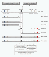
Summary of the results and definition of two domains in the structure of RAI1. By in silico analyses, several domains have been found for RAI1: a polyglutamine tract at the N-terminal of the protein (in blue), two polyserine domains (in yellow), a PHD domain at the C-terminal of RAI1 (in slanted lines) and two putative nuclear localization signals (in black). The schematic representation of all the mutants analyzed in this study is shown. An asterisk represents the missense mutations. Two defined domains are depicted.

Similar articles

Cited by

References

    1. Iafrate AJ, Feuk L, Rivera MN, Listewnik ML, Donahoe PK, Qi Y, Scherer SW, Lee C. Detection of large-scale variation in the human genome. Nat Genet. 2004;36:949. doi: 10.1038/ng1416. - DOI - PubMed
    1. Redon R, Ishikawa S, Fitch KR, Feuk L, Perry GH, Andrews TD, Fiegler H, Shapero MH, Carson AR, Chen W, Cho EK, Dallaire S, Freeman JL, González JR, Gratacòs M, Huang J, Kalaitzopoulos D, Komura D, MacDonald JR, Marshall CR, Mei R, Montgomery L, Nishimura K, Okamura K, Shen F, Somerville MJ, Tchinda J, Valsesia A, Woodwark C, Yang F. et al.Global variation in copy number in the human genome. Nature. 2006;444:444. doi: 10.1038/nature05329. - DOI - PMC - PubMed
    1. Beckmann JS, Estivill X, Antonarakis SE. Copy number variants and genetic traits: Closer to the resolution of phenotypic to genotypic variability. Nat Rev Genet. 2007;8:639. doi: 10.1038/nrg2149. - DOI - PubMed
    1. Korn JM, Kuruvilla FG, McCarroll SA, Wysoker A, Nemesh J, Cawley S, Hubbell E, Veitch J, Collins PJ, Darvishi K, Lee C, Nizzari MM, Gabriel SB, Purcell S, Daly MJ, Altshuler D. Integrated genotype calling and association analysis of SNPs, common copy number polymorphisms and rare CNVs. Nat Genet. 2008;40:1253. doi: 10.1038/ng.237. - DOI - PMC - PubMed
    1. McCarroll SA, Kuruvilla FG, Korn JM, Cawley S, Nemesh J, Wysoker A, Shapero MH, de Bakker PI, Maller JB, Kirby A, Elliott AL, Parkin M, Hubbell E, Webster T, Mei R, Veitch J, Collins PJ, Handsaker R, Lincoln S, Nizzari M, Blume J, Jones KW, Rava R, Daly MJ, Gabriel SB, Altshuler D. Integrated detection and population-genetic analysis of SNPs and copy number variation. Nat Genet. 2008;40:1166. doi: 10.1038/ng.238. - DOI - PubMed

Publication types