. 2010 Aug 25:10:259.
doi: 10.1186/1471-2148-10-259.

The evolutionary history of histone H3 suggests a deep eukaryotic root of chromatin modifying mechanisms

Affiliations

The evolutionary history of histone H3 suggests a deep eukaryotic root of chromatin modifying mechanisms

Jan Postberg et al. BMC Evol Biol. .

Abstract

Background: The phenotype of an organism is an outcome of both its genotype, encoding the primary sequence of proteins, and the developmental orchestration of gene expression. The substrate of gene expression in eukaryotes is the chromatin, whose fundamental units are nucleosomes composed of DNA wrapped around each two of the core histone types H2A, H2B, H3 and H4. Key regulatory steps involved in the determination of chromatin conformations are posttranslational modifications (PTM) at histone tails as well as the assembly of histone variants into nucleosomal arrays. Although the mechanistic background is fragmentary understood, it appears that the chromatin signature of metazoan cell types is inheritable over generations. Even less understood is the conservation of epigenetic mechanisms among eukaryotes and their origins.

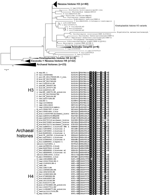
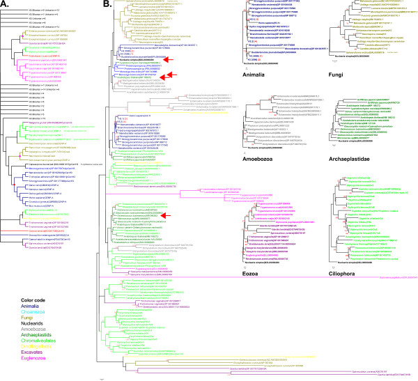
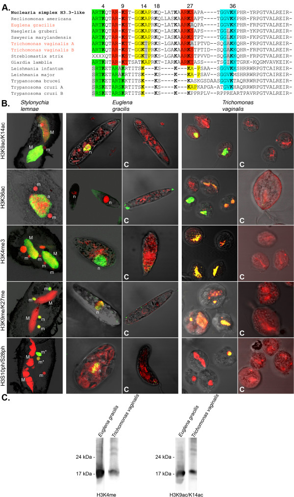
Results: In the light of recent progress in understanding the tree of eukaryotic life we discovered the origin of histone H3 by phylogenetic analyses of variants from all supergroups, which allowed the reconstruction of ancestral states. We found that H3 variants evolved frequently but independently within related species of almost all eukaryotic supergroups. Interestingly, we found all core histone types encoded in the genome of a basal dinoflagellate and H3 variants in two other species, although is was reported that dinoflagellate chromatin is not organized into nucleosomes.Most probably one or more animal/nuclearid H3.3-like variants gave rise to H3 variants of all opisthokonts (animals, choanozoa, fungi, nuclearids, Amoebozoa). H3.2 and H3.1 as well as H3.1t are derivatives of H3.3, whereas H3.2 evolved already in early branching animals, such as Trichoplax. H3.1 and H3.1t are probably restricted to mammals.We deduced a model for protoH3 of the last eukaryotic common ancestor (LECA) confirming a remarkable degree of sequence conservation in comparison to canonical human H3.1. We found evidence that multiple PTMs are conserved even in putatively early branching eukaryotic taxa (Euglenozoa/Excavata).

Conclusions: At least a basal repertoire of chromatin modifying mechanisms appears to share old common ancestry and may thus be inherent to all eukaryotes. We speculate that epigenetic principles responsive to environmental triggers may have had influenced phenotypic variation and concomitantly may potentially have had impact on eukaryotic diversification.


Figures

Figure 1
Figure 1
Phylogenetic relationsship between eukaryotic core histone types H3 and H4 as well as archaeal histones. A bootstrap consensus Neighbour Joining tree (A.) illustrates the phylogenetic relationsship between eukaryotic histones H3 and H4 as well as archaeal histones, which share common ancestry. More divergent H3 and H4 variants of kinetoplastids occur as sister groups with regard to their variants from other eukaryotes, probably due to long branch attraction. Similarly CenH3 variants occur as long branching sequences. The protein sequence alignment (B.) shows a conserved region from the histone fold domains of several eukaryotic histones H3 and H4 as well as archaeal histones. Residues identical in >95% of all sequences are shaded black. Residues similar in >95% of all sequences are shaded grey.
Figure 2
Figure 2
The evolutionary history of histone H3 and CenH3 variants. A. The evolutionary history of 159 H3 and CenH3 variants was inferred using the Neighbor-Joining method [48]. B. The evolutionary relationship of 128 non-redundant histone variants was inferred using the Neighbor-Joining method [48]. Importantly, animal stem H3 variants are identical in a broad range of species: For example, H3.3A96 (1) is identical in Trichoplax, Hydra, Nematostella, Buddenbrockia, and identical H3.3S96 (2) is found in Drosophila, Strongylocentrotus, Branchiostoma, Xenopus and many mammals. Further, H3.1 (3) is identical in mammals from mouse to human. Identical H3.2 (4) variants occur in organisms like Trichoplax, Drosophila, Branchiostoma, Xenopus and many mammals. The monophyly of several eukaryotic clades was well supported by phylogenetic analyses of histone H3 variant sequences. Pairwise comparison of selected H3 variants (indicated by arrows) from Unikonta or Bikonta species, respectively, revealed very high degrees of sequence conservation resulting in only rough separation of these clades. Due to very limited sequence variability no support for chromista or plant monophlyly could be found. However, two ciliate classes, Oligohymenophorea (e.g. Tetrahymena and Paramecium) and Spirotrichea (e.g. Stylonychia and Euplotes) were faithfully separated. Importantly, multiple H3 variants from Eozoa (Excavata + Euglenozoa) branched close to conserved H3 variants from other groups, predominantly Chromalveolata (Euglena, Reclinomonas, Sawyeria, Trichomonas, Streblomastix). All long branching Eozoa (Leishmania, Trypanosoma, Diplonema, Giardia, Spironucleus) or Microsporidia (Enterocytozoon, Encephalitozoon) H3 variants are parasites.
Figure 3
Figure 3
Numerous histone H3 variants are differentially expressed in the course of macronuclear differentiation in Stylonychia lemnae. A. Conservation of sequence motifs adjacent to N-terminal lysine residues and chaperone recognition sites in human H3.1 (top line) and Stylonychia histone H3 variants. The descending order of Stylonychia H3 variants reflects the phylogenetic distance compared to human H3.1. B. Agarose gel electrophoresis of PCR products amplified from developmental stage-specific cDNA. C. Quantitative Real Time PCR analysis of Stylonychia H3 variants expression. The morphology of developing macronuclei - revealed by confocal laser scanning microscopy of To-Pro-3 stained DNA - at successive stages is shown in relation to the time scale (hours post conjugation).
Figure 4
Figure 4
Reconstruction of ancestral histone H3 states. The ancestral state reconstruction of histone H3 variants from various clades (corresponding nodes are highlighted in the group-specific phylogenetic trees by red rhombs in Figure 2B) and H3 variant(s) of LECA confirms a high degree of sequence identity or similarity, respectively (shaded columns). Variable sites are highlighted (*); color scheme: basic amino acids (blue), acidic amino acids (red), aromatic amino acids (orange), putative phosphorylation sites (green). Nuclearia simplex H3 was used as outgroup for all group-specific trees. A detailed overview about the most frequent residues observed at such variable site is given in Additional file 4.
Figure 5
Figure 5
Multiple histone H3 modifications are conserved in putatively early branching eukaryotes. Posttranslational histone H3 modifications (PTM) occur at conserved N-terminal sequence motifs (color shaded in A.) in Euglena gracilis as well as Trichomonas vaginalis as suggested by immunofluorescence (B.) and Western (C.) analyses. C-marked images represent peptide competition assays as antibody specificity control. Both species represent putatively early branching eukaryotic clades. In the immunofluorescence panel (B.) the various PTMs occur as green signals, whereas nuclei and in some cases other nucleic acid-containing structures occur as red signals. In some cases DNA containing structures where labelled as follows: micronucleus/during mitosis (m/m*), macronucleus (M), nucleus (n). Western analyses (C.) confirm that the antibody targeted to H3K4me3 reacts with a protein band similar in size to histone H3 in Euglena and Trichomonas. Even H3K9ac/K14ac was detected in both Euglena and Trichomonas.

References

    1. Robinson PJ, Fairall L, Huynh VA, Rhodes D. EM measurements define the dimensions of the "30-nm" chromatin fiber: evidence for a compact, interdigitated structure. Proc Natl Acad Sci USA. 2006;103:6506–6511. doi: 10.1073/pnas.0601212103. - DOI - PMC - PubMed
    1. Tremethick DJ. Higher-order structures of chromatin: the elusive 30 nm fiber. Cell. 2007;128:651–654. doi: 10.1016/j.cell.2007.02.008. - DOI - PubMed
    1. Sandman K, Reeve JN. Chromosome packaging by archaeal histones. Adv Appl Microbiol. 2001;50:75–99. full_text. - PubMed
    1. Sandman K, Reeve JN. Structure and functional relationships of archaeal and eukaryal histones and nucleosomes. Arch Microbiol. 2000;173:165–169. doi: 10.1007/s002039900122. - DOI - PubMed
    1. Cubonova L, Sandman K, Hallam SJ, Delong EF, Reeve JN. Histones in crenarchaea. J Bacteriol. 2005;187:5482–5485. doi: 10.1128/JB.187.15.5482-5485.2005. - DOI - PMC - PubMed

Publication types

LinkOut - more resources