Review
. 2010 Oct;22(5):589-96.
doi: 10.1016/j.ceb.2010.07.012. Epub 2010 Aug 23.

Integration of planar cell polarity and ECM signaling in elongation of the vertebrate body plan

Affiliations
Review

Integration of planar cell polarity and ECM signaling in elongation of the vertebrate body plan

Paul Skoglund et al. Curr Opin Cell Biol. 2010 Oct.

Abstract

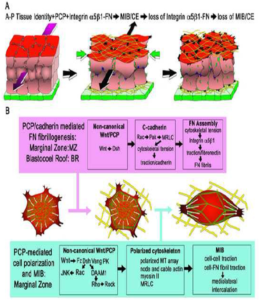
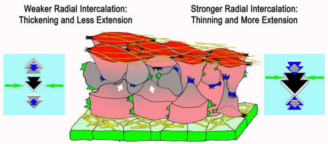
The shaping of the vertebrate embryonic body plan depends heavily on the narrowing and lengthening (convergence and extension) of embryonic tissues by cell intercalation, a process by which cells actively crawl between one another along the axis of convergence to produce a narrower, longer array. We discuss recent evidence that the vertebrate non-canonical Wnt/Planar Cell Polarity (PCP) pathway, known to directly function in polarizing the movements of intercalating cells, is also involved in the localized assembly of extracellular matrix (ECM). These cell-ECM interactions, in turn, are necessary for expression of the oriented, polarized cell intercalation. The mechanism of PCP/ECM interactions, their molecular signaling, and their mechanical consequences for morphogenesis are discussed with the goal of identifying important unsolved issues.


Figures

Figure 1
Figure 1
Diagrams show the deep mesodermal tissue of the marginal zone (red) in the process of undergoing convergence (green arrows) and extension (black arrow) by mediolateral cell intercalation (A). (The mediolateral axis is oriented across the page and the radial axis is vertical.) Initially unpolarized mesodermal cells (left) become polarized mediolaterally, in the plane of the tissue, and the resulting mediolaterally oriented traction drives the intercalation of cells along this axis (middle). Mediolaterally polarized protrusions occur at tissue surfaces bounding fibrillar FN (yellow), which are indicated as black protrusions, and internally, indicated by green protrusions. Polarization is dependent on anterior-posterior (A-P) tissue positional identity cues (magenta arrow), which aligns polarized behavior mediolateral to the A-P axis (see ref. 4). The expression of the polarized motility requires PCP signaling (see B, and refs. –7, 22) and FN-integrin α5β1 signaling (see B and refs. 8, 16). Absence of PCP signaling, or loss of FN-integrin signaling, results in loss of polarized behavior and failure of CE (right). The superficial endodermal epithelium (green) bounds the outside of marginal zone and reduces mesodermal surface tension, a necessary factor in allowing CE (26). The non-canonical Wnt/PCP pathway has dual roles in regulating polarized cell protrusive activity (B). PCP functions in a C-cadherin-mediated assembly of FN fibrils (yellow) in planarly unpolarized cells of blastocoel roof and in the planarly polarizing cells of the marginal zone (B, magenta box) (see refs. 9, 16, 21). This involves localization of actin (green) and development of adherens-like junctions at the cell periphery, and development of tensile forces at the cell periphery, which are necessary for fibrillogenesis (16). Expression of the polarized mediolateral intercalation behavior (MIB) (2–7, 31), which occurs only in the presumptive dorsal mesoderm of the marginal zone, is also dependent on integrin α5β1-FN signaling (B, blue box; see ref. 8). This involves mediolateral polarization the cytoskeleton, including formation of a mediolaterally oriented node-and-cable array of actin microfilaments, (green) (3), polarized microtubule growth (white arrows) (35), and mediolaterally polarized traction in the form of mediolaterally-directed protrusive activity (2).
Figure 2
Figure 2
A diagram shows a model of how cell-FN matrix interaction could regulate the amount of extension and thickening that results from convergence by regulating the forces generated by radial intercalation (blue arrows) and radial thickening forces (grey arrows) generated by compression of the tissue due to convergence (mediolateral intercalation, yellow arrows). Mediolaterally polarized protrusive activity at the tissue surfaces (black protrusions) and internally (green protrusions), drive mediolateral intercalation and generate convergence forces (yellow arrows), which brings the cells under compression and tends to bring about multi-layering and thickening (white arrows, grey cells). Radially polarized protrusive activity (blue protrusions) generates radial intercalation forces (blue arrows), which oppose the tendency to thicken. The balance of thickening forces and radial thinning forces determine the amount of thickening versus extension will result from convergence (see text, and refs. , –26,29).
Figure 3
Figure 3
Diagrams show the patterns of intercalation of notochord cells, and the amount of CE, in wild type embryos (A), a mutant of the PCP gene prickle (aimless, aim, B), a mutant of the laminin 3/4/5 subunit (chongmague, chm, C)and the double mutant (D) in the ascidian. Notochord cells are indicated with red, somites with yellow, and laminin with green. See refs. , , . Adapted with permission from ref. (issue 1, p. 39).
Figure 4
Figure 4
Diagrams of the vegetal aspect of the Xenopus embyo illustrates the mechanical function of the Fibrillin-2 (green) in reinforcement of notochordal (magenta)/somitic (yellow) boundary (NSB) in blastopore closure (A-B), concomitant with progressive dorsalization of posterior mesoderm (34, 36) (C). NSB formation proceeds progressively posteriorly in the mesoderm (green line, A-B) with the progress of MIB (transition of grey cells to red cells). Therefore large convergence forces (black arrows) develop across the newly formed NSB. Disruption of Fibrillin-2 results in fracture of this boundary and failure of blastopore closure (36) (B). Progression of MIB (red arrows) follows the earlier progress of nodal signaling (magenta arrows) and BMP inhibitors (blue arrows) into mesoderm of the ventral sector of the gastrula where these signals counter the effect of BMP signals (black arrows) and induce formation of posterior somitic mesoderm (C). The notochord and NSB, containing Fibrillin-2, potentially with BMP inhibiting activity, shears posteriorly along the lateral edge of the presumptive somitic mesoderm into ventral (presumptive posterior) regions (green arrows, C), where it may have dorsalizing activity. At the same time, it posterior movement has the mechanical effect of acting as a spline, a "zipper" that generates convergence forces as it pulls the pre-somitic mesoderm towards the midline (See 28, 34, 36, 40, 41; Dzamba, B. and DeSimone, D., personal communication).

Similar articles

Cited by

References

    1. Keller R, Davidson L, Edlund A, Elul T, Ezin M, Shook D, Skoglund P. Mechanisms of convergence and extension by cell intercalation. Philos Trans R Soc Lond B Biol Sci. 2000;355:897–922. - PMC - PubMed
    1. Shih J, Keller R. Cell motility driving mediolateral intercalation in explants of Xenopus laevis. Development. 1992;116:901–914. - PubMed
    1. Skoglund P, Rolo A, Chen X, Gumbiner BM, Keller R. Convergence and extension at gastrulation require a myosin IIB-dependent cortical actin network. Development. 2008;135:2435–2444. - PMC - PubMed
    1. Ninomiya H, Elinson RP, Winklbauer R. Antero-posterior tissue polarity links mesoderm convergent extension to axial patterning. Nature. 2004;430:364–367. - PubMed
    1. Wallingford JB, Rowning BA, Vogeli KM, Rothbacher U, Fraser SE, Harland RM. Dishevelled controls cell polarity during Xenopus gastrulation. Nature. 2000;405:81–85. - PubMed