. 2010 Oct;24(10):1904-16.
doi: 10.1210/me.2010-0175. Epub 2010 Aug 25.

The thyroid hormone receptor alpha1 protein is expressed in embryonic postmitotic neurons and persists in most adult neurons

Affiliations

The thyroid hormone receptor alpha1 protein is expressed in embryonic postmitotic neurons and persists in most adult neurons

Karin Wallis et al. Mol Endocrinol. 2010 Oct.

Abstract

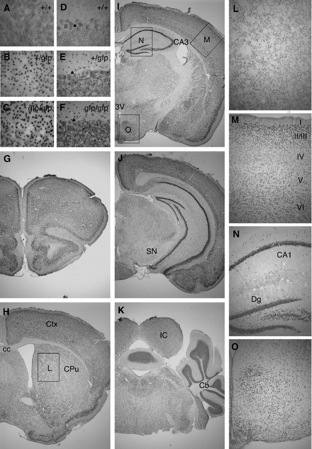
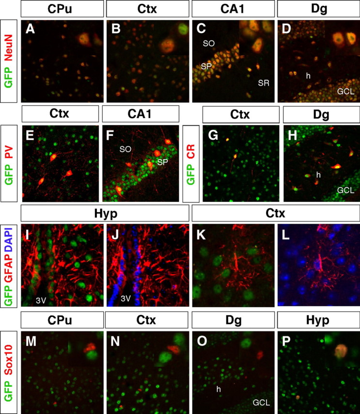
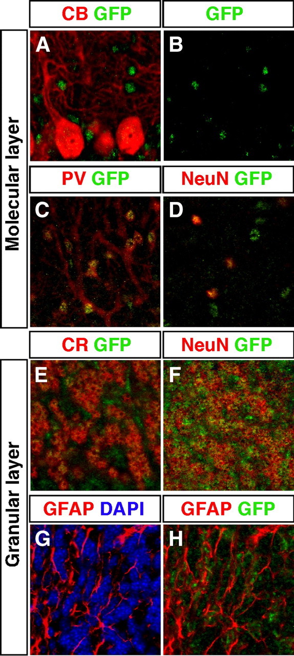
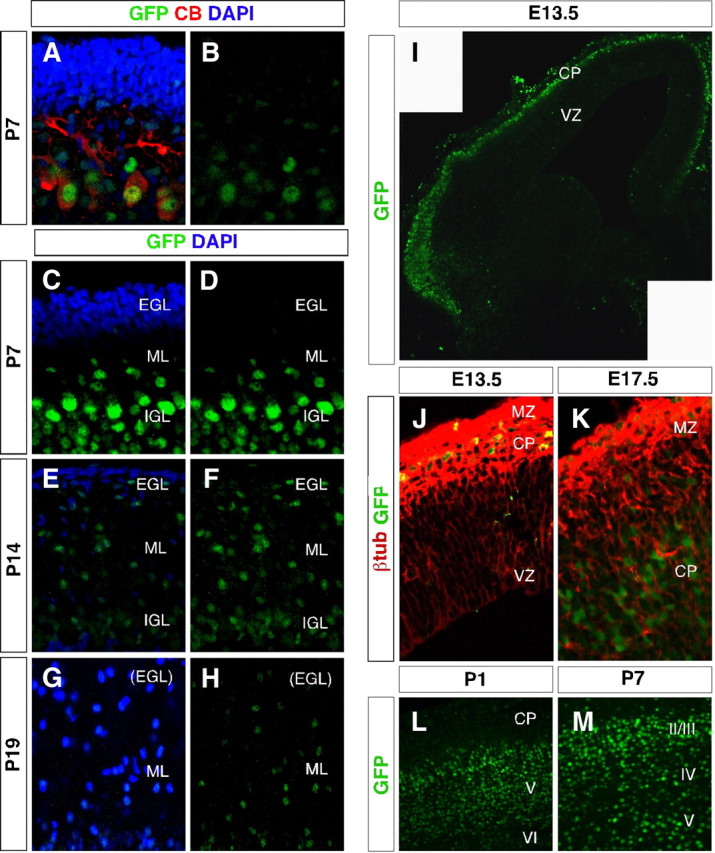
Thyroid hormone is essential for brain development where it acts mainly through the thyroid hormone receptor α1 (TRα1) isoform. However, the potential for the hormone to act in adult neurons has remained undefined due to difficulties in reliably determining the expression pattern of TR proteins in vivo. We therefore created a mouse strain that expresses TRα1 and green fluorescent protein as a chimeric protein from the Thra locus, allowing examination of TRα1 expression during fetal and postnatal development and in the adult. Furthermore, the use of antibodies against other markers enabled identification of TRα1 expression in subtypes of neurons and during specific stages of their maturation. TRα1 expression was first detected in postmitotic cells of the cortical plate in the embryonic telencephalon and preceded the expression of the mature neuronal protein NeuN. In the cerebellum, TRα1 expression was absent in proliferating cells of the external granular layer, but switched on as the cells migrated towards the internal granular layer. In addition, TRα1 was expressed transiently in developing Purkinje cells, but not in mature cells. Glial expression was found in tanycytes in the hypothalamus and in the cerebellum. In the adult brain, TRα1 expression was detected in essentially all neurons. Our data demonstrate that thyroid hormone, unexpectedly, has the capacity to play an important role in virtually all developing and adult neurons. Because the role of TRα1 in most neuronal cell types in vivo is largely unknown, our findings suggest that novel functions for thyroid hormone remain to be identified in the brain.


Figures

Fig. 1.
Fig. 1.
The knock-in TRα1-GFP allele. A, Map of the genomic locus encoding the TRα1 and -α2 isoforms and the rev-erbA gene, encoded by the opposing strand. Homologous recombination between the genomic locus and the targeting vector results in a targeted locus with GFP inserted in frame 3′ of exon 9 of the Thra1 gene. The resulting protein product is the chimeric TRα1-GFP protein. B, Correct homologous recombination was determined using Southern blot analyses of BamHI-cleaved genomic ES cell DNA hybridized with a 3′-probe that detect a sequence adjacent to but outside the targeting vector, or a GFP probe binding to the GFP sequence. The lane marked c is control DNA from heterozygote TRα1R384C mice. C, Southern blot analyses of BamHI-cleaved tail DNA, hybridized with a 5′-probe showing examples of all three genotypes after a heterozygote cross as well as heterozygote mice from which the neocassette had not been removed. D, Northern blot analyses of brain DNA from wt, homozygote, and heterozygote mice revealing that knock-in of GFP results in abolished expression from the Thra2 gene (2.6 kb). Mature TRα1-GFP RNA was 5.8 kb in size whereas the wt RNA was 5.2 kb. The insertion of GFP resulted in a novel 3.6-kb transcript in TRα1+/gfp and TRα1gfp/gfp mice and was detectable with all probes, indicating that it is a nonfunctional RNA. E, Quantitative real-time PCR showed the absence of TRα2 mRNA in homozygote mice and that expression was reduced to half in heterozygotes as compared with wt mice, whereas expression of TRα1 was increased in TRα1-GFP mice. F, Western blot of JEG-3 cell extracts after transfection with pCMV-TRα1, pCMV-TRα1-GFP; a nuclear extract from HeLa cells infected with a vaccinia virus vector expressing the chicken c-erbA/TRα protein is shown as a control. Lane c is a control with extract from untransfected cells. The membranes were hybridized with antibodies against TRα1 (left) or GFP and β-actin (right). TRα1: 46 kDa; TRα1-GFP: 75 kDa; β-actin: 42 kDa. G–I, Gene-regulatory functions of TRα1-GFP in transfected JEG-3 cells. Ligand-activated TRα1-GFP protein was equally potent as wt TRα1, as shown by detection of luciferase activity after cotransfection with 0, 50, or 150 ng of TRα1 and a reporter plasmid containing TRE-F2T2 (panel G) or TRE-Pal (H) response elements in front of a TK-luciferase cassette. The chimeric protein was not as effective in suppressing luciferase transcription as the wt protein, when cotransfected at 100 ng with increasing amounts of the corepressor NCoR in addition to the TRE-Pal reporter plasmid (I). All transfections were performed both in the presence and absence of 50 nm T3 in the medium and done in triplicate. *, P ≤ 0.05; **, P ≤ 0.01; ***, P ≤ 0.001, as compared with wt mice. DBD, DNA-binding domain; g, gfp; LBD, ligand-binding domain; UTR, untranslated region.
Fig. 2.
Fig. 2.
Physiological parameters of TRα1-GFP mice. Weight development of males (A) and females (B) from 3 d after birth until 6 wk of age demonstrated minor aberrancies in heterozygote but not in homozygote mice. Body weights of adult males (C) and females (D) showed no significant difference between the genotypes. E, Organ weights (grams per body weight) of male and female adult mice showed no alterations in brain or heart weights, whereas male homozygotes had significantly smaller livers as compared with wt controls. F and G, Serum total T4 (F) and T3 (G) levels of adult mice. Only a small difference was detected between the T3 levels of wt and heterozygote male mice; all other samples were normal. H, Analyses of relative gene expression levels of pituitary TSHβ, liver-specific Dio1, and cardiac MyHCα showed no differences between genotypes, whereas an increased expression of cardiac MyHCβ was seen in hearts of homozygous mutants. *, #, ‡, P ≤ 0.05; ##, P ≤ 0.01. *, wt vs. heterozygote; #, wt vs. homozygote; ‡, heterozygote vs. homozygote. TT3, total T3; TT4, total T4.
Fig. 3.
Fig. 3.
TRα1-GFP is expressed in gray matter areas of the adult brain. A–O, Micrographs of sections of the brain after immunohistochemistry for GFP to detect TRα1-GFP with DAB staining. Immunoreactivity was absent in wt mice (A and D) with higher expression in homozygotes (C and F) than in heterozygotes (B and H), as shown in the cortex (A–C) and in the cerebellum (D–F); magnification, ×400. The arrows in D–F indicate the Purkinje cells that were equally stained in all groups and thus only show background staining. G–K, ×20 magnification overviews. Boxes in H–J indicate ×100 magnifications in L–O. TRα1-GFP expression in the caudate putamen was structured and corresponds to gray matter areas (L). Immunoreactivity was detected in all cortical layers but was notably lower in layer IV (M). In the hippocampus TRα1-GFP was expressed in the stratum pyramidale and in the granular cell layer and hilar region of the dentate gyrus, as well as scattered in the stratum oriens and the stratum radiatum (N). In addition, a large number of cells in the hypothalamus expressed TRα1-GFP (O). cc, Corpus callosum; Cb, cerebellum; CPu, caudate putamen (striatum); Ctx, cortex; Dg, dentate gyrus; IC, inferior colliculus; SN, substantia nigra; 3V, third ventricle.
Fig. 4.
Fig. 4.
TRα1-GFP is expressed in virtually all neurons but in only a few specialized glial cells. A–P, Confocal imaging micrographs were taken of adult sections of the brain after immunohistochemistry against GFP to detect TRα1-GFP and markers for different cell types. Insets show high magnifications of the same region. A–D, TRα1-GFP expression was detected in all analyzed neurons (NeuN+ cells) in the adult brain, including in the caudate putamen (A), somatosensory cortex (B), CA1 region (C), and the dentate gyrus (D). E–H, Expression in GABAergic interneurons. TRα1-GFP was detected in PV+ interneurons in the cortex (E) and in the CA1 region of the hippocampus (F). TRα1-GFP and CR were coexpressed in interneurons of the cortex (G) and in interneurons and mossy cells in the hilus of the hippocampus (H). I–L, TRα1-GFP expression was detected in GFAP+ tanycytes lining the third ventricle of the hypothalamus (I and J), but not in astrocytes as shown in the cortex (K and L). Nuclei were counterstained with 4′6,-diamidino-2-phenylindole (DAPI). M–P, Double immunolabeling of oligodendrocytes with Sox10 show that TRα1-GFP was expressed only in adult oligodendrocytes of the hypothalamus (P), but not in the caudate putamen, cortex, or dentate gyrus (M–O). CPu, Caudate putamen; Ctx, cortex; GCL, granular cell layer; h, hilus; Hyp, hypothalamus; SO, stratum oriens; Sox, SRY related high-mobility group box; SP, stratum pyramidale; SR, stratum radiatum; 3V, third ventricle.
Fig. 5.
Fig. 5.
TRα1-GFP is expressed in specific neurons and glial cells in the cerebellum. A–H, Confocal imaging micrographs of adult sections showing expression of TRα1-GFP (detected with GFP antibody) and markers for different cell types. Examination of Purkinje cells marked with calbindin (CB) showed no colocalization with TRα1-GFP (A and B). In contrast, in the molecular layer, TRα1-GFP was expressed in PV+ stellate/basket cells (C) and in all neurons marked with NeuN (D). In the granular layer TRα1-GFP was expressed in granular cells as shown by immunostaining for both CR (E) and NeuN (F). TRα1-GFP was also expressed in GFAP+ glia cells (G and H). Nuclei were counterstained with 4′6,-diamidino-2-phenylindole (DAPI) in G.
Fig. 6.
Fig. 6.
TRα1-GFP is expressed in postmitotic neurons in the developing brain. A–M, Confocal imaging micrographs showing TRα1-GFP expression (detected with GFP antibody) in the developing cerebellum and telencephalon. A–H, Expression in the cerebellum. In contrast to adults, calbindin (CB)-positive Purkinje cells coexpressed TRα1-GFP at P7 (A and B). No TRα1-GFP expression was detected in the external granular layer at P7, P14, or P19, but in migrating cells of the molecular layer and in the internal granular layer (A–F). Nuclei were counterstained with 4′6,-diamidino-2-phenylindole (DAPI) (A, C, E, and G). I–M, Expression in the telencephalon. In the E13.5 telencephalon, TRα1-GFP was expressed in the marginal zone and cortical plate, but not in the ventricular zone (I and J). The image (panel I) is a montage of two micrographs. The TRα1-GFP-positive cells expressed the postmitotic neuronal marker β-tubIII (J). At E17.5 TRα1-GFP expression was higher in the cortical plate than in the marginal zone (K), whereas at P1 the expression was highest in the deeper cortical layers V and VI where the oldest neurons are located (L). At P7, TRα1-GFP was equally expressed in all cortical layers (M). CP, cortical plate; IGL, intergranular layer; ML, molecular layer; MZ, marginal zone; VZ, ventricular zone.

References

    1. Delange FM1996. Endemic cretinism. In: Braverman LE, Utiger RD eds. Werner and Ingbar’s the thyroid, 7th ed. Philadelphia: Lippincott-Raven Publishers; 756–767
    1. DeLong GR1996. The neuromuscular system and brain in hypothyroidism. In: Braverman LE, Utiger RD, eds. Werner and Ingbar’s the thyroid, 7th ed. Philadelphia: Lippincott-Raven Publishers; 826–835
    1. Morreale de Escobar G, Obregon MJ, Escobar del Rey F2004. Role of thyroid hormone during early brain development. Eur J Endocrinol 151(Suppl 3):U25–U37 - PubMed
    1. Sap J, Muñoz A, Damm K, Goldberg Y, Ghysdael J, Leutz A, Beug H, Vennström B1986. The c-erb-A protein is a high-affinity receptor for thyroid hormone. Nature 324:635–640 - PubMed
    1. Weinberger C, Thompson CC, Ong ES, Lebo R, Gruol DJ, Evans RM1986. The c-erb-A gene encodes a thyroid hormone receptor. Nature 324:641–646 - PubMed

Publication types

Substances