

doi: 10.1128/JVI.01133-10.
Epub 2010 Aug 25.
Environmental sources of scrapie prions
Affiliations
- PMID: 20739536
- PMCID: PMC2953180
- DOI: 10.1128/JVI.01133-10
Item in Clipboard
Environmental sources of scrapie prions
J Virol.
2010 Nov.
Abstract
Ovine scrapie and cervine chronic wasting disease show considerable horizontal transmission. Here we report that a scrapie-affected sheep farm has a widespread environmental contamination with prions. Prions were amplified by protein-misfolding cyclic amplification (sPMCA) from seven of nine environmental swab samples taken, including those from metal, plastic, and wooden surfaces. Sheep had been removed from the areas from which the swabs were taken up to 20 days prior to sampling, indicating that prions persist for at least that long. These data implicate inanimate objects as environmental reservoirs for prion infectivity that are likely to contribute to facile disease transmission.
Figures
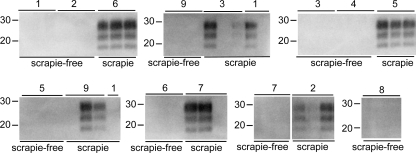
Amplification of prions from environmental samples. Prions were extracted from swabs taken of surfaces from a scrapie-free farm or a farm where scrapie is endemic. Swabs were taken from a wooden post (1), a plastic scratching post (2), and the following metal surfaces: fencing (3), gate (4 and 6), pen (5), feed trough (7), and water trough (8 and 9). Samples 1, 2, 3, 4, and 8 were taken from outdoor surfaces and 5, 6, 7, and 9 from indoor surfaces. Extracts were used as seeds for 8 rounds of sPMCA. Products were digested with proteinase K and analyzed by Western blotting. PrP was detected with monoclonal antibodies SHA31 and P4. Molecular-weight markers are shown.
References
-
- Castilla, J., P. Saá, and C. Soto. 2005. Detection of prions in blood. Nat. Med. 11:982-985. - PubMed
-
- Jones, M., A. H. Peden, C. V. Prowse, A. Gröner, J. C. Manson, M. L. Turner, J. W. Ironside, I. R. MacGregor, and M. W. Head. 2007. In vitro amplification and detection of variant Creutzfeldt-Jakob disease PrPSc. J. Pathol. 213:21-26. - PubMed
Publication types
MeSH terms
Substances
LinkOut - more resources
Full Text Sources
Other Literature Sources
