Comparative Study
. 2010 Aug 25;30(34):11288-99.
doi: 10.1523/JNEUROSCI.1764-10.2010.

In vivo NMDA receptor activation accelerates motor unit maturation, protects spinal motor neurons, and enhances SMN2 gene expression in severe spinal muscular atrophy mice

Affiliations
Comparative Study

In vivo NMDA receptor activation accelerates motor unit maturation, protects spinal motor neurons, and enhances SMN2 gene expression in severe spinal muscular atrophy mice

Olivier Biondi et al. J Neurosci. .

Abstract

Spinal muscular atrophy (SMA), a lethal neurodegenerative disease that occurs in childhood, is caused by the misexpression of the survival of motor neuron (SMN) protein in motor neurons. It is still unclear whether activating motor units in SMA corrects the delay in the postnatal maturation of the motor unit resulting in an enhanced neuroprotection. In the present work, we demonstrate that an adequate NMDA receptor activation in a type 2 SMA mouse model significantly accelerated motor unit postnatal maturation, counteracted apoptosis in the spinal cord, and induced a marked increase of SMN expression resulting from a modification of SMN2 gene transcription pattern. These beneficial effects were dependent on the level of NMDA receptor activation since a treatment with high doses of NMDA led to an acceleration of the motor unit maturation but favored the apoptotic process and decreased SMN expression. In addition, these results suggest that the NMDA-induced acceleration of motor unit postnatal maturation occurred independently of SMN. The NMDA receptor activating treatment strongly extended the life span in two different mouse models of severe SMA. The analysis of the intracellular signaling cascade that lay downstream the activated NMDA receptor revealed an unexpected reactivation of the CaMKII/AKT/CREB (cAMP response element-binding protein) pathway that induced an enhanced SMN expression. Therefore, pharmacological activation of spinal NMDA receptors could constitute a useful strategy for both increasing SMN expression and limiting motor neuron death in SMA spinal cord.


Figures
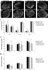
Figure 1.
Figure 1.
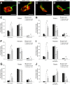
Effect of NMDA treatment on the muscle phenotype of type 2 SMA-like mice. A–H, Immunodetection of developmental MyHC isoforms (i.e., embryonic and neonatal) in the soleus of vehicle (A, E)- and NMDA-treated control mice (B, F) and vehicle (C, G)- and NMDA-treated type 2 SMA-like mice (D, H) at 12 d of age. Scale bar, 200 μm. I, Analysis of the embryonic (Emb) and neonatal (Neo) typology of three muscles of the calf (i.e., the soleus, the plantaris, and the tibialis muscles) from vehicle- and NMDA-treated control mice and type 2 SMA-like mice (n = 6), at 12 d of age. *p < 0.05; **p < 0.01. J, Determination of the cross-section area (CSA) of the soleus, the plantaris, and the tibialis from vehicle- and NMDA-treated control and type 2 SMA-like mice (n = 6) at 12 d of age. K, Number of muscle fibers in the soleus, plantaris, and tibialis muscles from vehicle- and NMDA-treated control and type 2 SMA-like mice (n = 6) at 12 d of age. Error bars indicate SD.
Figure 2.
Figure 2.
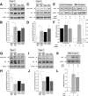
Effect of NMDA treatment on neuromuscular junction maturation in type 2 SMA-like mice. A–D, Motor endplate labeling with α-bungarotoxin, anti-neurofilament, and anti-synaptophysin antibodies in the soleus of vehicle (A)- and NMDA-treated control mice (B) and vehicle (C)- and NMDA-treated type 2 SMA-like mice (D) at 12 d of age showing a typical pretzel (B), perforated (A, D), and immature (C) type of NMJ. Scale bar, 20 μm. E–G, Analysis of the neuromuscular junction maturation in the soleus, the plantaris, and the tibialis muscles from vehicle- and NMDA-treated control and type 2 SMA-like mice (n = 5) at 12 d of age. H–J, Determination of the neuromuscular junction area in the soleus, plantaris, and tibialis from vehicle- and NMDA-treated control and type 2 SMA-like mice (n = 5) at 12 d of age. *p < 0.05; **p < 0.01. Error bars indicate SD.
Figure 3.
Figure 3.
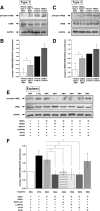
Neuroprotective effect of NMDA in type 2 SMA-like mice. A–D, Immunodetection of ChAT-positive motor neurons in the lumbar spinal cord (L1–L5) of vehicle (A)- and NMDA-treated control mice (B), and vehicle (C)- and NMDA-treated type 2 SMA-like mice (D) at 12 d of age. Scale bar, 100 μm. E, F, Quantitative analysis of the number (E) and the cell body area (F) of motor neurons per ventral horn in the lumbar spinal cord of vehicle-treated control and type 2 SMA-like mice (n = 6) compared with NMDA-treated control and type 2 SMA-like mice (n = 6) at 12 d of age. G, H, Western blot analysis and quantification of cleaved caspase 3 protein expression in the ventral lumbar spinal cord of vehicle- and NMDA-treated control and type 2 SMA-like mice at 12 d of age (n = 3). *p < 0.05; **p < 0.01. Error bars indicate SD.
Figure 4.
Figure 4.
Effect of NMDA on muscle function, life span, and weight curve in SMA-like mice. A, Picture showing NMDA-treated type 2 SMA-like mice during a grip test on a metal rod at 12 d of age. B, Grip time of vehicle-, and NMDA-treated type 2 SMA-like mice compared with vehicle-treated controls (n = 13, n = 11, and n = 15, respectively). C, Behavior of NMDA-treated type 2 SMA-like mice during an open field test at 12 d of age. D, Total number of crossings during 5 min for the NMDA-treated (n = 13) compared with vehicle-treated type 2 SMA-like mice (n = 11). E, Number of peripheral crossings for the NMDA-treated mice compared with the vehicle-treated mice. F, Life span of NMDA-treated (n = 30) compared with vehicle-treated type 2 SMA-like mice (n = 30). G, Phenotype of control mice (Control) compared with vehicle-treated type 2 SMA-like mice (Vehicle SMA) and NMDA-treated type 2 SMA-like mice (NMDA SMA) at 12 d of age. H, Weight curve in NMDA-treated (n = 20) compared with vehicle-treated type 2 SMA-like mice (n = 20). I, Life span of NMDA-treated (n = 25) compared with vehicle-treated type 1 SMA-like mice (n = 25). J, Weight curve in NMDA-treated (n = 15) compared with vehicle-treated type 1 SMA-like mice (n = 15) and vehicle-treated control (n = 15). *p < 0.05; **p < 0.01; ***p < 0.001. Error bars indicate SD.
Figure 5.
Figure 5.
SMN expression in the spinal cord of vehicle- and NMDA-treated SMA-like mice. A, B, Western blot analysis and quantification of SMN protein expression in the ventral lumbar spinal cord of control mice at 6 d of age, vehicle type 1 SMA-like mice at 2 d of age, and NMDA-treated type 1 SMA-like mice at 6 d of age (n = 3). C, D, Western blot analysis and quantification of SMN protein expression in the ventral lumbar spinal cord of control mice, vehicle- and NMDA-treated type 2 SMA-like mice at 12 d of age (n = 4). E, F, Quantification by real-time RT-PCR of exon 7–exon 8 (E7–E8) segment-containing SMN transcripts normalized to 18S transcripts in the ventral lumbar spinal cord of vehicle type 1 and type 2 SMA-like mice at 2 and 12 d of age, respectively, and of NMDA-treated type 1 and type 2 SMA-like mice at 6 and 12 d of age, respectively (E). Quantification by real-time RT-PCR of exon 7–exon 8 (E7–E8) segment-containing SMN transcripts normalized to exon 4–exon 5 (E4–E5) segment-containing SMN transcripts in the ventral lumbar spinal cord of vehicle type 1 and type 2 SMA-like mice at 2 and 12 d of age, respectively, and of NMDA-treated type 1 and type 2 SMA-like mice at 6 and 12 d of age, respectively (F). G, RT-PCR analysis of SMN full-length transcripts (SMN FL, 392 nt) and SMN transcripts lacking exon 7 (SMNΔ7, 338 nt) in the ventral lumbar spinal cord of vehicle type 1 SMA-like mice at 2 d of age and NMDA-treated type 1 SMA-like mice at 6 d of age. H, RT-PCR analysis of SMN full-length transcripts (SMN FL, 392 nt) and SMN transcripts lacking exon 7 (SMNΔ7, 338 nt) in the ventral lumbar spinal cord of vehicle- and NMDA-treated type 2 SMA-like mice at 12 d of age. *p < 0.05; **p < 0.01. Error bars indicate SD.
Figure 6.
Figure 6.
Evaluation of the role of the Ca2+ signaling in NMDA-induced SMN expression in SMA spinal cord and SMA explants. A, B, Labeling of neuromuscular junctions with α-bungarotoxin, anti-neurofilament, and anti-synaptophysin antibodies in control (A) and SMA (B) spinal cord explants showing similar structures. Scale bar, 20 μm. C, D, Western blot analysis and quantification of SMN protein expression in control and SMA spinal cord explants in presence or not of NMDA, MK801, and calcium chelators EGTA and BAPTA-AM (n = 3). E, F, Western blot analysis and quantification of CaMKII protein phosphorylation in the ventral lumbar spinal cord of control mice at 6 d of age, of vehicle type 1 SMA-like mice at 2 d of age, and NMDA-treated type 1 SMA-like mice at 6 d of age (n = 3). G, H, Western blot analysis and quantification of CaMKII protein phosphorylation in the ventral lumbar spinal cord of control mice, vehicle- and NMDA-treated type 2 SMA-like mice at 12 d of age (n = 3). I, J, Western blot analysis and quantification of SMN protein expression in SMA spinal cord explants in presence or not of NMDA and CaMKII inhibitor KN-93 (n = 3). *p < 0.05. Error bars indicate SD.
Figure 7.
Figure 7.
Evaluation of AKT and ERK phosphorylation in NMDA-induced SMN expression in SMA spinal cord and SMA explants. A, B, Western blot analysis and quantification of AKT protein phosphorylation in the ventral lumbar spinal cord of control mice at 6 d of age, of vehicle type 1 SMA-like mice at 2 d of age, and NMDA-treated type 1 SMA-like mice at 6 d of age (n = 3). C, D, Western blot analysis and quantification of AKT protein phosphorylation in the ventral lumbar spinal cord of control mice, vehicle- and NMDA-treated type 2 SMA-like mice at 12 d of age (n = 3). E, F, Western blot analysis and quantification of SMN protein expression in the control and SMA spinal cord explants in presence or not of NMDA and PI3K inhibitor LY294002 (n = 3). G, H, Western blot analysis and quantification of ERK protein phosphorylation in the ventral lumbar spinal cord of control mice at 6 d of age, of vehicle type 1 SMA-like mice at 2 d of age, and NMDA-treated type 1 SMA-like mice at 6 d of age (n = 3). I, J, Western blot analysis and quantification of ERK protein phosphorylation in the ventral lumbar spinal cord of control mice, vehicle- and NMDA-treated type 2 SMA-like mice at 12 d of age (n = 3). K, L, Western blot analysis and quantification of SMN protein expression in SMA spinal cord explants in presence or not of NMDA and ERK inhibitor U0126 (n = 3). *p < 0.05. Error bars indicate SD.
Figure 8.
Figure 8.
Evaluation of CREB phosphorylation in NMDA-induced SMN expression in SMA spinal cord and SMA explants. A, B, Western blot analysis and quantification of CREB protein phosphorylation in the ventral lumbar spinal cord of control mice at 6 d of age, of vehicle type 1 SMA-like mice at 2 d of age, and NMDA-treated type 1 SMA-like mice at 6 d of age (n = 3). C, D, Western blot analysis and quantification of CREB protein phosphorylation in the ventral lumbar spinal cord of control mice, vehicle- and NMDA-treated type 2 SMA-like mice at 12 d of age (n = 3). E, F, Western blot analysis and quantification of CREB protein phosphorylation in the control and SMA spinal cord explants in presence or not of NMDA, MK801, BAPTA-AM, PI3K inhibitor LY294002, CaMKII inhibitor KN-93, and ERK inhibitor U0126 (n = 3). *p < 0.05. Error bars indicate SD.

References

    1. Allemand E, Batsché E, Muchardt C. Splicing, transcription, and chromatin: a ménage à trois. Curr Opin Genet Dev. 2008;18:145–151. - PubMed
    1. Andreassi C, Patrizi AL, Monani UR, Burghes AH, Brahe C, Eboli ML. Expression of the survival of motor-neuron (SMN) gene in primary neurons and increase in SMN levels by activation of the N-methyl-d-aspartate glutamate receptor. Neurogenetics. 2002;4:29–36. - PubMed
    1. Biondi O, Grondard C, Lécolle S, Deforges S, Pariset C, Lopes P, Cifuentes-Diaz C, Li H, della Gaspera B, Chanoine C, Charbonnier F. Exercise-induced activation of NMDA receptor promotes motor unit development and survival in a type 2 spinal muscular atrophy model mouse. J Neurosci. 2008;28:953–962. - PMC - PubMed
    1. Brunet A, Datta SR, Greenberg ME. Transcription-dependent and -independent control of neuronal survival by the PI3K-Akt signaling pathway. Curr Opin Neurobiol. 2001;11:297–305. - PubMed
    1. Cartaud A, Strochlic L, Guerra M, Blanchard B, Lambergeon M, Krejci E, Cartaud J, Legay C. MuSK is required for anchoring acetylcholinesterase at the neuromuscular junction. J Cell Biol. 2004;165:505–515. - PMC - PubMed

Publication types

MeSH terms

Substances