. 2010 Nov;104(5):2863-72.
doi: 10.1152/jn.00253.2010. Epub 2010 Aug 25.

Multiple somatotopic representations of heat and mechanical pain in the operculo-insular cortex: a high-resolution fMRI study

Affiliations

Multiple somatotopic representations of heat and mechanical pain in the operculo-insular cortex: a high-resolution fMRI study

Ulf Baumgärtner et al. J Neurophysiol. 2010 Nov.

Abstract

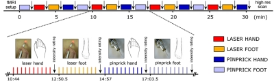
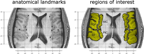
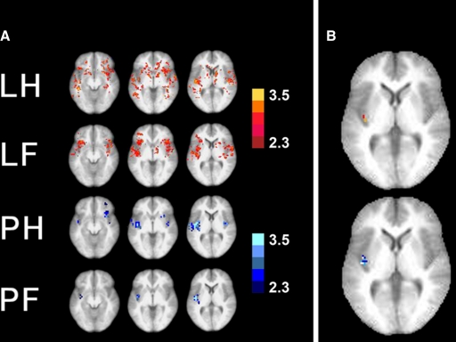
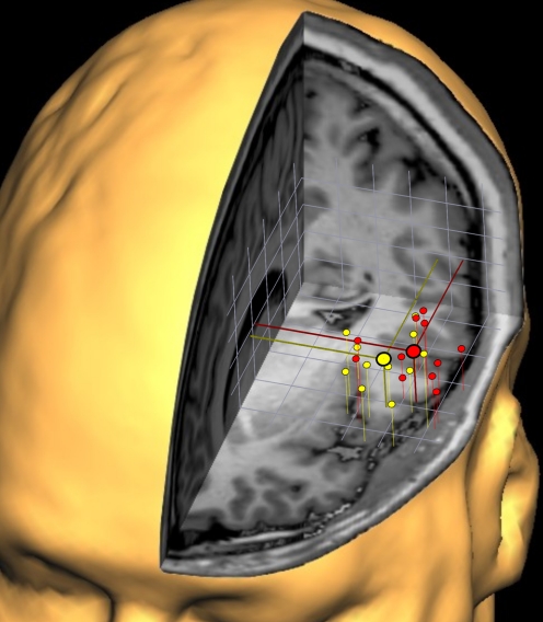
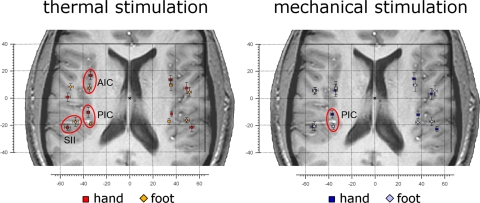
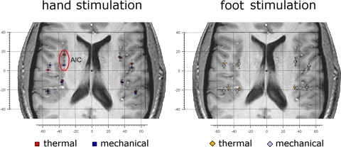
Whereas studies of somatotopic representation of touch have been useful to distinguish multiple somatosensory areas within primary (SI) and secondary (SII) somatosensory cortex regions, no such analysis exists for the representation of pain across nociceptive modalities. Here we investigated somatotopy in the operculo-insular cortex with noxious heat and pinprick stimuli in 11 healthy subjects using high-resolution (2 × 2 × 4 mm) 3T functional magnetic resonance imaging (fMRI). Heat stimuli (delivered using a laser) and pinprick stimuli (delivered using a punctate probe) were directed to the dorsum of the right hand and foot in a balanced design. Locations of the peak fMRI responses were compared between stimulation sites (hand vs. foot) and modalities (heat vs. pinprick) within four bilateral regions of interest: anterior and posterior insula and frontal and parietal operculum. Importantly, all analyses were performed on individual, non-normalized fMRI images. For heat stimuli, we found hand-foot somatotopy in the contralateral anterior and posterior insula [hand, 9 ± 10 (SD) mm anterior to foot, P < 0.05] and in the contralateral parietal operculum (SII; hand, 7 ± 10 mm lateral to foot, P < 0.05). For pinprick stimuli, we also found somatotopy in the contralateral posterior insula (hand, 9 ± 10 mm anterior to foot, P < 0.05). Furthermore, the response to heat stimulation of the hand was 11 ± 12 mm anterior to the response to pinprick stimulation of the hand in the contralateral (left) anterior insula (P < 0.05). These results indicate the existence of multiple somatotopic representations for pain within the operculo-insular region in humans, possibly reflecting its importance as a sensory-integration site that directs emotional responses and behavior appropriately depending on the body site being injured.


Figures

Fig. 1.
Fig. 1.
Experimental design. The figure illustrates the timing of actions performed during the functional magnetic resonance imaging (fMRI) experiment. Laser heat and pinprick stimuli were delivered to the skin of the dorsum of the hand and of the foot by an experimenter inside the scanner room. Stimuli of the same modality and to the same body site were delivered in blocks of 10, with an interstimulus interval of 11.5 s. At the end of each block, the subject was asked to provide an average intensity rating for that block. The order of blocks was balanced across subjects.
Fig. 2.
Fig. 2.
Variability in structural anatomy and definition of regions of interest on EPI scans. Left: the variability in structural anatomy of the population explored in this study. The individual brains were aligned to the anterior commissure (AC, origin of axes) to display variability of structural anatomy of the insular and opercular regions on a single brain. As AC is approximately located in the center of the brain as well as of the regions of interest (ROIs), this Talairach-like alignment was chosen to minimize the influence of interindividual anatomical differences. Each dot represents the average location of the landmarks used to define the eight ROIs. On each hemisphere, the medial dots indicate the anterior pole of the insula, the middle of the curvature of the insula, the sulcus between the 3rd and 4th insular gyri (the anatomical separation of anterior and posterior insula) (Bense et al. 2001) and the posterior pole of the insula. The lateral dot indicates the location where the central sulcus ends (separation between the frontal and the parietal operculum). The single dot on the midline represents the posterior commissure (PC). Axis scaling is in mm, error bars represent the standard error of the mean. Right: the manner in which 8 ROIs (in yellow) were determined in a representative subject. FOP, frontal operculum; AIC, anterior insular cortex; PIC, posterior insular cortex; SII, secondary somatosensory cortex.
Fig. 3.
Fig. 3.
Group analysis. A: at the significance threshold of Z = 2.3, 3 transversal slices through the operculo-insular region are shown (left to right; 4 mm below AC level, 4 mm above AC level, 12 mm above AC level) in 4 lines for the 4 modalities (laser hand, laser foot, pinprick hand, pinprick foot). Although especially for the laser stimuli, the brain activations are scattered throughout the operculo-insular and frontal cortices, the brain area that is activated most consistently across the 4 different stimulation modalities is the left posterior insula. B: brain activation of both hand and foot stimulation is shown on the same slice (laser: top; pinprick: bottom). At an elevated threshold of Z = 3.8, a differential somatotopic representation within the posterior insula in anterior-posterior direction can be seen: the hand representation is anterior to the foot representation. Top: laser hand (red pixels) vs. foot (orange pixels). Bottom: pinprick hand (dark blue) vs. foot (light blue), overlapping pixels in white. Note that on group analysis level, the only somatotopic representation that can be identified is in the posterior insula. Left: the left side of the brains.
Fig. 4.
Fig. 4.
Three-dimensional clustering of the individual cortical responses obtained within SII following laser hand (red) and foot (yellow) stimulation. To gain an impression of the variability of cortical representations within SII, the individual activations of all subjects (n = 11) were projected onto 1 subject's 3-dimensional reconstruction of the brain. The individual coordinate systems were aligned to the anterior commissure and the AC-PC plane. Despite the overlap between the scatterings of the red and yellow symbols, a systematic difference in medial-lateral direction is visible, and the average localizations for hand and foot representation are illustrated by the large symbols. Grid width: 1 cm.
Fig. 5.
Fig. 5.
Somatotopic representation of hand and foot following noxious stimulation. Left: the average location of the activations in response to heat (laser) stimulation of the hand (red squares) and foot dorsum (orange diamonds) in each ROI. As indicated by red ellipsoids, the location of the activations was significantly different (circled in red) in the contralateral anterior insula, posterior insula, and parietal operculum (SII; for a quantitative description of location differences see Table 1A). Right: the average location of the activations in response to pinprick (punctate probe) stimulation of the hand (dark blue squares) and foot dorsum (pale blue diamonds), in each ROI. The location of the activations was significantly different in the contralateral posterior insula (for a quantitative description of location differences see Table 1B). In all panels, error bars represent SE. The left side of the brains is shown on the left. AIC, anterior insular cortex; PIC, posterior insular cortex; SII, secondary somatosensory cortex.
Fig. 6.
Fig. 6.
Different location of activations in response to heat and pinprick stimulation. Left: the average location of the activations in response to heat (laser) and pinprick (punctate probe) stimulation of the hand dorsum in each ROI. Red squares represent heat stimulation, blue squares represent pinprick stimulation. The location of the activations was significantly different in the contralateral anterior insula (circled in red; for a quantitative description of location differences see Table 2A). Right: the average location of the activations in response to heat (laser) and pinprick (punctuate probe) stimulation of the foot dorsum. Orange diamonds represent heat stimulation, pale blue diamonds represent pinprick stimulation. For a quantitative description of location differences see Table 2B. In all panels error bars represent SE. The left side of the brains is shown on the left. AIC, anterior insular cortex.

References

    1. Afif A, Hoffmann D, Becq G, Guenot M, Magnin M, Mertens P. MRI-based definition of a stereotactic two-dimensional template of the human insula. Stereotact Funct Neurosurg 87: 385–394, 2009 - PubMed
    1. Afif A, Hoffmann D, Minotti L, Benabid AL, Kahane P. Middle short gyrus of the insula implicated in pain processing. Pain 138: 546–555, 2008 - PubMed
    1. Albanese MC, Duerden EG, Rainville P, Duncan GH. Memory traces of pain in human cortex. J Neurosci 27: 4612–4620, 2007 - PMC - PubMed
    1. Andersson JLR, Lilja A, Hartvig P, Långström B, Gordh T, Handwerker H, Torebjörk E. Somatotopic organization along the central sulcus, for pain localization in humans, as revealed by positron emission tomography. Exp Brain Res 117: 192–199, 1997 - PubMed
    1. Apkarian AV, Bushnell C, Treede RD, Zubieta JK. Human brain mechanisms of pain perception and regulation in health and disease. Eur J Pain 9: 463–484, 2005 - PubMed

Publication types