. 2010 Dec;88(6):1241-9.
doi: 10.1189/jlb.0610332. Epub 2010 Aug 25.

PD-L1(hi) retinal pigment epithelium (RPE) cells elicited by inflammatory cytokines induce regulatory activity in uveitogenic T cells

Affiliations

PD-L1(hi) retinal pigment epithelium (RPE) cells elicited by inflammatory cytokines induce regulatory activity in uveitogenic T cells

Yan Ke et al. J Leukoc Biol. 2010 Dec.

Abstract

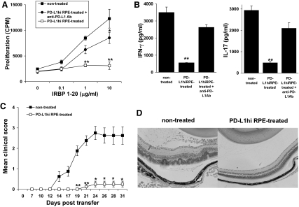
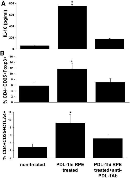
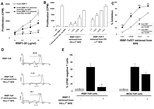
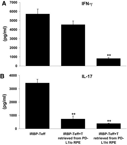
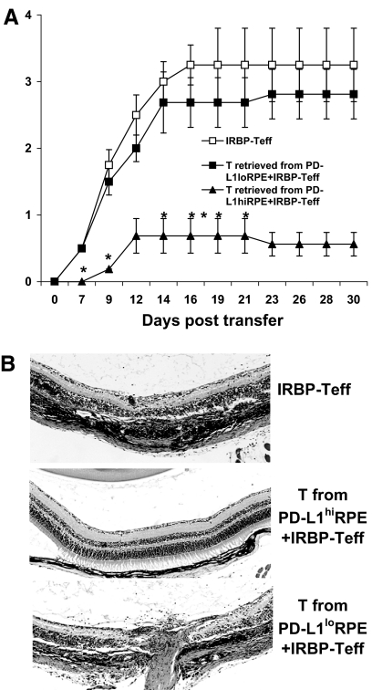
We previously reported that after exposure to inflammatory cytokines, such as IL-17 and IFN-γ, RPE cells express increased amounts of suppressor of cytokine signaling, leading to general suppression of the inflammatory response. Here, we demonstrate that RPE cells expressed increased levels of PD-L1 in response to IL-17, IFN-γ, or Poly I:C. These PD-L1(hi) RPE cells inhibited the pathogenic activities of IRBP-specific T cells, which usually induced uveitis when injected into naïve mice (EAU). The suppressed pathogenicity of these uveitogenic T cells after exposure to PD-L1(hi) RPE cells could be partially reversed by anti-PD-L1 antibodies. Nevertheless, IRBP-specific T cells pre-exposed to PD-L1(hi) RPE cells displayed substantial suppressor activity, which strongly inhibited the activation of fresh IRBP-Teffs in response to subsequent antigenic challenge and when transferred into naïve mice, inhibited the induction of EAU by IRBP-Teff transfer. These findings suggest that inflammatory cytokine-triggered up-regulation of PD-L1 on RPE constitutes a critical factor for inducing infiltrated uveitogenic T cells with regulatory activities, which may accelerate the natural resolution of T cell-mediated intraocular inflammation.


Figures

Figure 1.
Figure 1.. PD-L1 expression on RPE cells.
(A) PD-L1 expression is up-regulated on RPE cells but not RACs in response to inflammatory stimuli. RPE and RACs were cultured to confluence and then were incubated for 24 h with medium alone, 500 U/ml IFN-γ, 100 ng/ml IL-17, or 50 μg/ml Poly I:C, and PD-L1 expression was measured by flow cytometry (thick line: isotype control; thin line: anti-PD-L1-FITC). (B–D) The increase in PD-L1 expression on RPE cells caused by IL-17 is dose-dependent. (B) RPE cells were cultured for 24 h in medium (dotted line) or medium containing 1 ng/ml (dark, solid line) or 100 ng/ml (bold, solid line) of IL-17, and then PD-L1 expression was examined. The light, solid line is a cell stained with the isotype control antibody. (C) Analysis of PD-L1 expression after culture of RPE with increasing doses of IL-17. (D) Analysis of PD-L1 expression on a per-cell basis, quantified via MFI. (E) Kinetic analysis of PD-L1 expression by RPE cells in response to IL-17. RPE cells were cultured with 100 ng/ml IL-17, as in A, for 6–48 h, and then PD-L1 expression on RPE cells was measured by flow cytometry (left panel). After 24 h culture with IL-17, cells were washed and recultured in fresh culture medium without IL-17 for 6–48 h, and then PD-L1 expression on RPE cells was examined (right panel). The white columns are untreated cells. The results of one experiment with triplicate cultures are shown and are representative of the results for three separate. Error bars are the sd. *P < 0.05 versus medium without IL-17.
Figure 2.
Figure 2.. IRBP-specific T cells lose pathogenic activity after incubation with PD-L1hi RPE cells.
(A) IRBP-specific T cells (5×10⁶) were left untreated or were preincubated with monolayers of PD-L1hi RPE cells (ratio of 10:1), with or without anti-PD-L1 mAb (20 μg/ml) for 24 h, and then, the recovered T cells were cocultured with the indicated concentration of IRBP1–20 peptide and irradiated, syngeneic, splenic APCs and their proliferation measured after 48 h. (B) Cells were treated as in A but using 10 μg/ml IRBP1–20 for stimulation, and then (B), the IFN-γ or IL-17 released by the IRBP-T cells was measured and (C) their uveitis-inducing ability after adoptive transfer (5×10⁶/mouse; n=6 of two experiments) evaluated. (D) Pathological examination of the eye in the two groups performed on Day 31. **P < 0.01 and *P < 0.05 versus nontreated.
Figure 3.
Figure 3.. IRBP-specific T cells preincubated with PD-L1hi RPE acquire the Treg phenotype.
Cells were treated as in Fig. 2B, and then, the IL-10 produced by the IRBP-T cells was measured by ELISA (A), and CD4/CD25/Foxp3- or CTLA4-positive cells were detected by three-color antibody staining, followed by flow cytometry analysis (B). *P < 0.05 versus nontreated.
Figure 4.
Figure 4.. IRBP-T cells retrieved from the preincubation with PD-L1hi RPE potently inhibit IRBP-Teff proliferation.
IRBP-specific T cells (5×10⁶) were preincubated with PD-L1hi or PD-L1lo RPE cells for 24 h, retrieved (named IRBP-T), and added to fresh IRBP-specific T cells (IRBP-Teff) at a ratio of 1:1 in the presence of indicated doses of IRBP1–20 and irradiated APCs (A) or at a different ratio in the presence of 10 μg/ml IRBP1–20 (B). After 48 h, the proliferation of the IRBP-Teffs in response to antigen stimulation was evaluated. (C) Percentage inhibition of the proliferation of fresh IRBP-Teffs by retrieved IRBP-T cells from the incubation with PD-L1hi or PD-L1lo RPE cells calculated from B. (D) IRBP-T cells retrieved from PD-L1hi or PD-L1lo RPE cell monolayers were tested for suppressor activity as in A. Prior to the assay, fresh T cells were stained with CFSE. The numbers indicate the percentage of CFSE-negative cells analyzed by flow cytometry. Results of one experiment, representative of three experiments, are shown. FL1-H, Fluorescence 1-height. (E) Fresh IRBP-Teffs or MOG-Teffs, prestained with CFSE, were incubated with IRBP1–20 or MOG35–55, as appropriate, and irradiated APCs in the presence (+) or absence (–) of IRBP-T cells retrieved from PD-Llhi RPE cells and then CFSE-negative cells analyzed by flow cytometry. The columns indicate the mean percentage of CFSE-negative cells after 5 days of culture. The error bars indicate the sd for the experimental results for three consecutively tested donor/responder pairs. The light-gray columns show the percent of CFSE-negative, fresh T cells without IRBP or MOG antigen stimulation Results shown are representative of those from three independent experiments. (A, B, E) **P < 0.01 and *P < 0.05 versus fresh IRBP-Teff; (C) **P < 0.01 and *P < 0.05 versus fresh IRBP-T: T from PD-L1lo RPE.
Figure 5.
Figure 5.. IRBP-T cells retrieved from the preincubation with PD-L1hi RPE cells reduce IFN-γ produced by IRBP-Teffs.
IRBP-specific T cells (5×10⁶) were preincubated with PD-L1hi or PD-L1lo RPE cells for 24 h, retrieved (named IRBP-T), and added to fresh, IRBP-specific T cells (IRBP-Teff) at a ratio of 1:1 in the presence of 10 μg/ml IRBP1–20 and irradiated APCs. After 48 h, the production of Th1 and Th17 cytokines (A and B) by IRBP-Teffs, in response to antigen stimulation, was evaluated by ELISA. **P < 0.01 versus IRBP-Teff.
Figure 6.
Figure 6.. IRBP-T cells retrieved from the preincubation with PD-L1hi RPE cells suppress EAU induced by IRBP-Teffs.
Three groups of naïve mice (n=6; two experiments) were adoptively transferred with blast IRBP-Teffs (5×10⁶/mouse) after in vitro stimulation with IRBP1–20 and irradiated APCs for 2 days. Group 1 mice received only IRBP-Teffs (positive control). Groups 2 and 3 were injected with IRBP-T cells after incubation with PD-L1hi or PD-L1lo RPE cells, respectively (experimental groups). Disease was monitored and scored clinically (A) two times/week and histologically (B) at Day 30. *P < 0.05 versus IRBP-Teff.

References

    1. Luger D., Silver P. B., Tang J., Cua D., Chen Z., Iwakura Y., Bowman E. P., Sgambellone N. M., Chan C. C., Caspi R. R. (2008) Either a Th17 or a Th1 effector response can drive autoimmunity: conditions of disease induction affect dominant effector category. J. Exp. Med. 205, 799–810 - PMC - PubMed
    1. Peng Y., Han G., Shao H., Wang Y., Kaplan H. J., Sun D. (2007) Characterization of IL-17+ interphotoreceptor retinoid-binding protein-specific T cells in experimental autoimmune uveitis. Invest. Ophthalmol. Vis. Sci. 48, 4153–4161 - PMC - PubMed
    1. Amadi-Obi A., Yu C. R., Liu X., Mahdi R. M., Clarke G. L., Nussenblatt R. B., Gery I., Lee Y. S., Egwuagu C. E. (2007) TH17 cells contribute to uveitis and scleritis and are expanded by IL-2 and inhibited by IL-27/STAT1. Nat. Med. 13, 711–718 - PubMed
    1. Cox C. A., Shi G., Yin H., Vistica B. P., Wawrousek E. F., Chan C. C., Gery I. (2008) Both Th1 and Th17 are immunopathogenic but differ in other key biological activities. J. Immunol. 180, 7414–7422 - PMC - PubMed
    1. Ishida K., Panjwani N., Cao Z., Streilein J. W. (2003) Participation of pigment epithelium in ocular immune privilege. 3. Epithelia cultured from iris, ciliary body, and retina suppress T-cell activation by partially non-overlapping mechanisms. Ocul. Immunol. Inflamm. 11, 91–105 - PubMed

Publication types