. 2010 Nov;299(5):C1079-90.
doi: 10.1152/ajpcell.00548.2009. Epub 2010 Aug 25.

Orai1 interacts with STIM1 and mediates capacitative Ca2+ entry in mouse pulmonary arterial smooth muscle cells

Affiliations

Orai1 interacts with STIM1 and mediates capacitative Ca2+ entry in mouse pulmonary arterial smooth muscle cells

Lih Chyuan Ng et al. Am J Physiol Cell Physiol. 2010 Nov.

Abstract

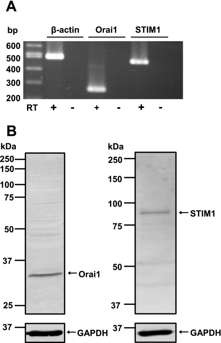
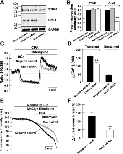
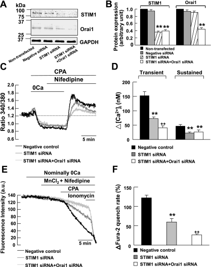
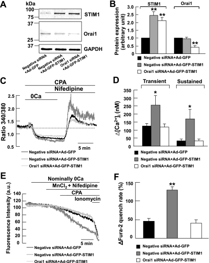
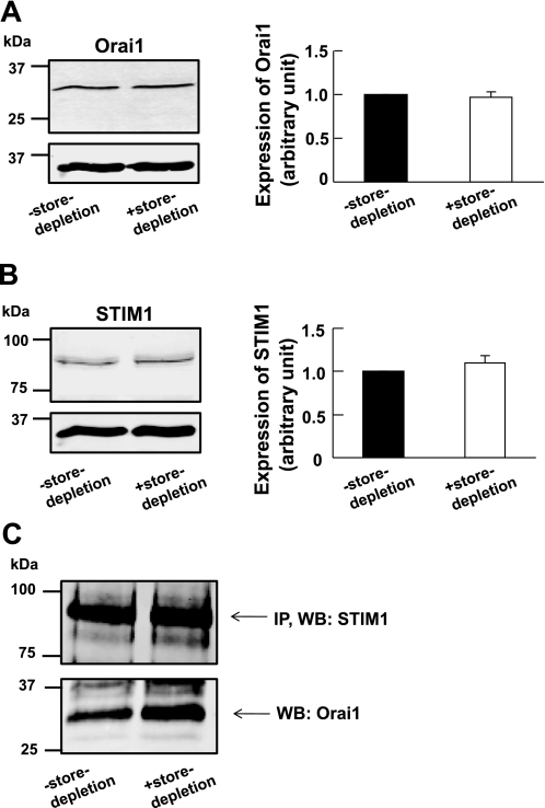
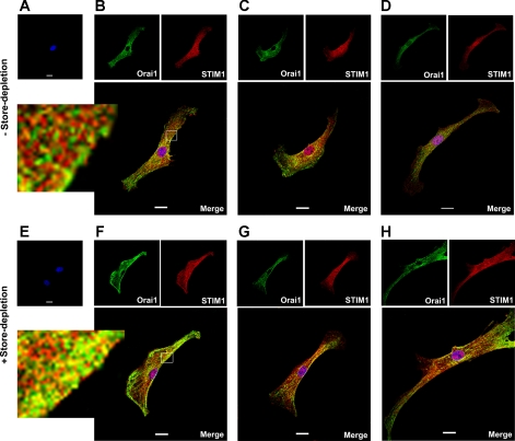
Previous studies in mouse pulmonary arterial smooth muscle cells (PASMCs) showed that cannonical transient receptor potential channel TRPC1 and stromal interaction molecule 1 (STIM1) mediate the sustained component of capacitative Ca(2+) entry (CCE), but the molecular candidate(s) that mediate the transient component of CCE remain unknown. The aim of the present study was to examine whether Orai1 mediates the transient component of CCE through activation of STIM1 in mouse PASMCs. In primary cultured mouse PASMCs loaded with fura-2, cyclopiazonic acid (CPA) caused a transient followed by a sustained rise in intracellular Ca(2+) concentration ([Ca(2+)](i)). The transient but not the sustained rise in [Ca(2+)](i) was partially inhibited by nifedipine. The nifedipine-insensitive transient rise in [Ca(2+)](i) and the increase in Mn(2+) quench of fura-2 fluorescence caused by CPA were both reduced in cells treated with Orai1 siRNA. These responses to CPA were further reduced in cells treated with Orai1 and STIM1 small interfering (si)RNA. Moreover, overexpression of STIM1 enhanced the rise in [Ca(2+)](i) and the increase in Mn(2+) quench of fura-2 fluorescence caused by CPA, and these responses were reduced in cells treated with Orai1 siRNA. RT-PCR revealed Orai1 and STIM1 mRNAs, and Western blot analysis identified Orai1 and STIM1 proteins in mouse PASMCs. Furthermore, Orai1 was found to coimmunoprecipitate with STIM1, and the precipitation level of Orai1 was increased in cells subjected to store-depletion. Immunostaining revealed colocalization of Orai1 and STIM1 proteins, and the colocalization of these proteins was more apparent after store-depletion. These data provide direct evidence that the transient component of CCE is mediated by Orai1 channel as a result of STIM1 activation in mouse PASMCs.


Figures

Fig. 1.
Fig. 1.
Orai1 and stromal-interacting molecule 1 (STIM1) expression in mouse pulmonary arterial smooth muscle cells (PASMCs). A: RT-PCR products from cultured mouse PASMCs amplified using primers for mouse Orai1 (269 bp), STIM1 (473 bp), and β-actin (498 bp). Three separate RT-PCR reactions were performed in the presence (+) and absence (−) of reverse transcriptase (RT). B: Orai1, STIM1, and GAPDH proteins were detected in cultured mouse PASMCs using Western blot analysis. Experiments were performed in 5 separate Western blot analyses.
Fig. 2.
Fig. 2.
Orai1 mediates capacitative Ca2+ entry (CCE) in mouse PASMCs. A and B: Orai1 protein and GAPDH were detected in nontransfected mouse PASMCs and in PASMCs transfected with 200 nM scrambled small interfering RNA (siRNA; negative control). The expression of Orai1 but not GAPDH was reduced significantly in cells transfected with 200 nM Orai1 siRNA. Experiments were performed in 3 separate Western blot analyses (**P < 0.01, ANOVA). C: siRNA knockdown of Orai1 reduced the cyclopiazonic acid (CPA)-induced transient but not the sustained increase in fura-2 fluorescence ratio in the presence of 10 μM nifedipine. 0Ca, Ca2+-free solution. D: bar graph showing mean changes in transient and sustained increase in intracellular Ca2+ concentration ([Ca2+]i) caused by 10 μM CPA after readdition of 2 mM Ca2+ in the presence of 10 μM nifedipine, in negative control cells (filled bars, n = 89), and in Orai1 siRNA-transfected cells (open bars, n = 86). **P < 0.01 (unpaired t-test). E: siRNA knockdown of Orai1 reduced the increase in Mn2+ quench of fura-2 fluorescence caused by 10 μM CPA in the presence of 10 μM nifedipine. AU, arbitrary units. F: bar graph showing percentage change in fura-2 quench rate after store-depletion in the presence of 10 μM nifedipine, in negative control cells (filled bar, n = 79), and in Orai1 siRNA-transfected cells (open bar, n = 86). **P < 0.01 (unpaired t-test).
Fig. 3.
Fig. 3.
STIM1 is associated with Orai1 to mediate CCE in mouse PASMCs. A and B: STIM1, Orai1, and GAPDH proteins were detected in nontransfected mouse PASMCs and in PASMCs transfected with 200 nM scrambled siRNA (negative control). The expression of STIM1 but not Orai1 or GAPDH was reduced significantly in cells transfected with 200 nM STIM1 siRNA. The expressions of STIM1 and Orai1 but not GAPDH were reduced significantly in cells transfected with both 200 nM STIM1 siRNA and 200 nM Orai1 siRNA. Experiments were performed in 3 separate Western blot analyses (**P < 0.01, ANOVA). C: siRNA knockdown of STIM1 reduced the CPA-induced transient and sustained increase in fura-2 fluorescence ratio in the presence of 10 μM nifedipine. siRNA knockdown of STIM1 and Orai1 further reduced the CPA-induced transient but not sustained increase in fura-2 fluorescence ratio in the presence of nifedipine. D: bar graph showing mean changes in transient and sustained increase in [Ca2+]i caused by 10 μM CPA after readdition of 2 mM Ca2+ in the presence of 10 μM nifedipine, in negative control cells (filled bars, n = 103), in STIM1 siRNA-transfected cells (shaded bars, n = 148), and in STIM1 siRNA and Orai1 siRNA-transfected cells (open bars, n = 70). **P < 0.01, compared with negative control cells (ANOVA); ++P < 0.01, compared with negative control cells and STIM1 siRNA-transfected cells (ANOVA). E: siRNA knockdown of STIM1 reduced the increase in Mn2+ quench of fura-2 fluorescence caused by 10 μM CPA in the presence of 10 μM nifedipine. siRNA knockdown of STIM1 and Orai1 further reduced the increase in Mn2+ quench of fura-2 fluorescence caused by CPA in the presence of nifedipine. F: bar graph showing percentage change in fura-2 quench rate after store-depletion in the presence of 10 μM nifedipine, in negative control cells (filled bar, n = 125), in STIM1 siRNA-transfected cells (shaded bar, n = 151), and in STIM1 siRNA and Orai1 siRNA-transfected cells (open bar, n = 137). **P < 0.01, compared with negative control cells (ANOVA); ++P < 0.01, compared with the negative control cells and STIM1 siRNA-transfected cells (ANOVA).
Fig. 4.
Fig. 4.
knockdown of Orai1 reduced CCE in STIM1-overexpressing mouse PASMCs. A and B: STIM1, Orai1, and GAPDH proteins were detected in cells infected with adenovirus containing green fluorescent protein (Ad-GFP) transfected with 200 nM scrambled siRNA. The expression of STIM1 but not Orai1 or GAPDH increased markedly in cells infected with STIM1-GFP-adenovirus (Ad-GFP-STIM1) transfected with scrambled siRNA. The expression of Orai1 but not STIM1 or GAPDH was reduced significantly in STIM1-overexpressing cells transfected with 200 nM Orai1 siRNA. Experiments were performed in 3 separate Western blot analyses (**P < 0.01, ANOVA). C: overexpression of STIM1 in scrambled siRNA-transfected cells caused an increase in CPA-induced transient and sustained rise in fura-2 fluorescence ratio in the presence of 10 μM nifedipine. The increases in fluorescence ratio were reduced in Orai1 siRNA-transfected cells overexpressed with STIM1. D: bar graph showing mean changes in transient and sustained increase in [Ca2+]i caused by 10 μM CPA after readdition of 2 mM Ca2+ in the presence of 10 μM nifedipine, in GFP-infected cells transfected with scrambled siRNA (filled bars, n = 33), in STIM1-overexpressing cells transfected with scrambled siRNA (shaded bars, n = 65), and in STIM1-overexpressing cells transfected with Orai1 siRNA (open bars, n = 50). *P < 0.05, compared with GFP-infected cells transfected with scrambled siRNA and STIM1-overexpressing cells transfected with Orai1 siRNA (ANOVA). E: overexpression of STIM1 in scrambled siRNA-transfected cells caused a CPA-induced increase in Mn2+ quench of fura-2 fluorescence in the presence of 10 μM nifedipine. The increases in Mn2+ quench of fura-2 fluorescence was reduced in Orai1 siRNA-transfected cells overexpressed with STIM1. F: bar graph showing percentage change in fura-2 quench rate after store-depletion in the presence of 10 μM nifedipine, in GFP-infected cells transfected with scrambled siRNA (filled bars, n = 40), in STIM1-overexpressing cells transfected with scrambled siRNA (shaded bars, n = 95), and in STIM1-overexpressing cells transfected with Orai1 siRNA (open bars, n = 57). **P < 0.01, compared with GFP-infected cells transfected with scrambled siRNA and STIM1-overexpressing cells transfected with Orai1 siRNA (ANOVA).
Fig. 5.
Fig. 5.
Orai1 coimmunoprecipitates with STIM1 in mouse PASMCs. A, left: Orai1 was detected in cultured mouse PASMCs in the absence and presence of store-depletion. Right: bar graph showing expression levels of Orai1 measured relative to GAPDH in control cells (denoted as 1, filled bar) and in cells subjected to store-depletion (open bar). Data are means ± SE of 7 separate Western blot analyses. B, left: STIM1 was detected in cultured mouse PASMCs in the absence and presence of store-depletion. Right: bar graph showing expression levels of STIM1 measured relative to GAPDH in control cells (denoted as 1, filled bar) and in cells subjected to store-depletion (open bar). Data are means ± SE of 7 separate Western blot analyses. C: STIM1 coimmunoprecipitated Orai1 in cultured mouse PASMCs in the absence and presence of store-depletion. STIM1 was first immunoprecipitated (IP) with EXBIO STIM1 antibody (10 μg), and the blot was subsequently probed with BD Biosciences STIM1 antibody (WB, 1:100). The blot was then probed for coimmunoprecipitation (co-IP) of Orai1 expression using Orai1 antibody (WB, 1:100, ProSci). Experiments were performed in 3 separate co-IP procedures and Western blot analyses.
Fig. 6.
Fig. 6.
Colocalization of Orai1 and STIM1 in mouse PASMCs. A–D: staining of mouse cultured PASMCs after exposure of live cells with normal bath physiological salt solution (PSS). A: omission of Orai1 and STIM1 antibody resulted in no Orai1 or STIM1 staining. B–D: three representative cells dual-labeled with anti-Orai1 antibody (green) and STIM1 antibody (red). Orai1 and STIM1 colocalization (yellow/orange) is shown in the merged images. E–H: staining of mouse cultured PASMCs after exposure of live cells with Ca2+-free PSS containing 10 μM CPA. E: omission of Orai1 and STIM1 antibody resulted in no Orai1 or STIM1 staining. F–H: three representative cells dual-labeled with anti-Orai1 antibody (green) and STIM1 antibody (red). Colocalization of Orai1 and STIM1 is more apparent (yellow/orange) after store-depletion as shown in the merged images. Nuclei were stained with DAPI (blue). Experiments were performed in 3 separate immunostaining procedure, each with duplicate coverslips. Scale bars, 20 μm.

References

    1. Albert AP, Large WA. Activation of store-operated channels by noradrenaline via protein kinase C in rabbit portal vein myocytes. J Physiol 544: 113–125, 2002 - PMC - PubMed
    1. Albert AP, Large WA. Store-operated Ca2+-permeable non-selective cation channels in smooth muscle cells. Cell Calcium 33: 345–356, 2003 - PubMed
    1. Albert AP, Saleh SN, Peppiatt-Wildman CM, Large WA. Multiple activation mechanisms of store-operated TRPC channels in smooth muscle cells. J Physiol 583: 25–36, 2007 - PMC - PubMed
    1. Baryshnikov SG, Pulina MV, Zulian A, Linde CI, Golovina VA. Orai1, a critical component of store-operated Ca2+ entry, is functionally associated with Na+/Ca2+ exchanger and plasma membrane Ca2+ pump in proliferating human arterial myocytes. Am J Physiol Cell Physiol 297: C1103–C1112, 2009 - PMC - PubMed
    1. Berra-Romani R, Mazzocco-Spezzia A, Pulina MV, Golovina VA. Ca2+ handling is altered when arterial myocytes progress from a contractile to a proliferative phenotype in culture. Am J Physiol Cell Physiol 295: C779–C790, 2008 - PMC - PubMed

Publication types

MeSH terms

LinkOut - more resources