. 2010 Sep;120(9):3120-6.
doi: 10.1172/JCI43267. Epub 2010 Aug 25.

Induced pluripotent stem cell-derived hepatocytes have the functional and proliferative capabilities needed for liver regeneration in mice

Affiliations

Induced pluripotent stem cell-derived hepatocytes have the functional and proliferative capabilities needed for liver regeneration in mice

Silvia Espejel et al. J Clin Invest. 2010 Sep.

Abstract

The ability to generate induced pluripotent stem (iPS) cells from a patient's somatic cells has provided a foundation for organ regeneration without the need for immune suppression. However, it has not been established that the differentiated progeny of iPS cells can effectively reverse failure of a vital organ. Here, we examined whether iPS cell-derived hepatocytes have both the functional and proliferative capabilities needed for liver regeneration in mice with fumarylacetoacetate hydrolase deficiency. To avoid biases resulting from random genomic integration, we used iPS cells generated without viruses. To exclude compensation by hepatocytes not derived from iPS cells, we generated chimeric mice in which all hepatocytes were iPS cell derived. In vivo analyses showed that iPS cells were intrinsically able to differentiate into fully mature hepatocytes that provided full liver function. The iPS cell-derived hepatocytes also replicated the unique proliferative capabilities of normal hepatocytes and were able to regenerate the liver after transplantation and two-thirds partial hepatectomy. Thus, our results establish the feasibility of using iPS cells generated in a clinically acceptable fashion for rapid and stable liver regeneration.


Figures
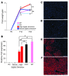
Figure 1
Figure 1. iPS cell–derived hepatocytes facilitate NTBC-independent growth and survival of FAH-deficient mice.
(A) Relative postnatal weight gain in chimeric mice compared with wild-type and FAH-deficient (KO) controls. Chimeric mice were separated into 2 groups, based on high versus no/low contribution of iPS cells to genomic DNA from digits (Supplemental Figure 1). NTBC withdrawal was initiated on P6. In contrast to KO controls and mice with no/low levels of digital chimerism, mice with high levels of digital chimerism did not require reinstatement of NTBC treatment after P22 for growth and survival. All control mice were on the 129S4 strain background, while both iPS cells and blastocysts were on a mixed 129S4 and C57BL/6 background. C57BL/6 neonates grow faster, which explains the slight difference in weight gain between control and chimeric mice between P6 and P19. (B) Quantitative RT-PCR shows that growth and survival in the absence of NTBC are strictly correlated with Fah expression in the liver. Liver Fah expression is detectable in all mice with a high iPS cell contribution to digits but not in mice with no/low levels of digital chimerism. Fah expression approaches wild-type levels with time off NTBC, increasing from approximately 50% (range, 33%–69%) in mice analyzed at P28 (left red bar) to approximately 80% (range, 76%–78%) in mice analyzed at P70 (right red bar). Maximum Fah expression levels in iPS cell chimeric mice are similar to levels found after approximately 100% liver repopulation in mice generated by injection of normal ES cells into FAH-deficient blastocysts (ESCs). (CF) FAH immunostaining (red). Representative liver sections show that mice with (C) no or (D) low digital chimerism lack FAH-expressing cells. Livers of mice with high levels of digital chimerism show repopulation with FAH-positive cells, between approximately (E) 50% at P28 and (F) 100% at P70. Nuclei are stained blue. Scale bars: 100 μm. Data represent mean ± SEM. *P < 0.05; **P < 0.005; #P > 0.05.
Figure 2
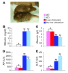
Figure 2. Restoration of liver function in FAH-deficient mice repopulated with iPS cell–derived hepatocytes.
(A) In the absence of NTBC, a mouse with no digital chimerism is lethargic and shows signs of dehydration and lack of grooming (front), while a littermate with high levels of digital chimerism appears normal (back) at P22. (BE) Blood analysis of liver function parameters at P22 in mice deprived of NTBC since P6. (B) Bilirubin and (C) albumin levels reflect the detoxification and protein synthesis functions of the liver, respectively. (D) Alkaline phosphatase (AP) is a marker of cholestasis, (E) while alanine aminotransferase (ALT) indicates hepatocyte injury. Mice with iPS cell contribution to the liver (all mice with high levels of digital chimerism, referred to as liver chimerism) have values indistinguishable from wild-type mice (including undetectable bilirubin), while the results from mice with no liver chimerism (all mice with no/low levels of digital chimerism) and FAH-deficient mice indicate liver failure. Liver function parameters of the mice with liver chimerism were stable at reanalysis at P70 and P300 (Supplemental Figure 2). Data represent mean ± SEM. **P < 0.005; #P > 0.05.
Figure 3
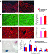
Figure 3. iPS cell–derived hepatocytes have normal proliferative capabilities.
(A) FAH immunostaining (red, both images) shows approximately 90% liver repopulation with FAH-expressing hepatocytes in a chimeric mouse at P28. Additional Ki67 immunostaining (green, right image) shows proliferating FAH-positive hepatocytes bordering growth-arrested FAH-negative hepatocytes. (B and C) Analysis of liver regeneration after two-thirds partial hepatectomy in 3 wild-type and 3 FAH-deficient mice repopulated to approximately 100% with transplanted iPS cell–derived hepatocytes (FAH immunostaining, green or red). (B) BrdU labeling (red) 40 hours after two-thirds partial hepatectomy shows that timing and magnitude of DNA synthesis are indistinguishable between wild-type hepatocytes (left) and iPS cell–derived hepatocytes (middle). Quantification of BrdU-labeled hepatocytes in all mice (right). (C) Immunostaining for phosphorylated histone H3 (p-hH3, green) 48 hours after two-thirds partial hepatectomy shows that wild-type hepatocytes (left) and iPS cell–derived hepatocytes (middle) progress into mitosis with similar efficiency. Quantification of p-hH3–labeled hepatocytes in all mice (right). (D) Combined FAH immunostaining (red) and X-gal staining (blue) shows 2 nodules of similar size derived from a single iPS cell–derived hepatocyte or a Rosa26 hepatocyte transplanted together into an immune-deficient, FAH-deficient mouse. The blue X-gal staining masks the red FAH signal. Both FAH immunostaining and X-gal staining are highly sensitive and specific (Figure 1, E and F, and Supplemental Figure 6). Classifying repopulating nodules based on the number of hepatocytes visible in 2-dimensional liver sections (cells per nodule) indicates that cotransplanted Rosa26 hepatocytes and iPS cell–derived hepatocytes are equally effective in clonal expansion. The number of cell divisions required for formation of repopulating nodules composed of 11–25, 26–50, 51–75, 76–100, or 101–125 hepatocytes is 7, 8, 9, 10, or 11, respectively. This calculation is based on the assumption that each hepatocyte in the nodule proliferates to the same extent and that nodules are 3-dimensional spheres (35). Results of 3 transplantation experiments are shown. Nuclei are stained blue. Scale bars: 100 μm (A, left image, and D); 50 μm (B and C); 25 μm (A, right image). Data represent mean ± SEM. #P > 0.05.
Figure 4
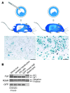
Figure 4. Differentiation of iPS cells into hepatocytes occurs in the absence of cell fusion.
(A) Use of a ubiquitously and conditionally expressed genetic marker to exclude that iPS cells acquire hepatocyte function by fusion with blastocyst-derived hepatocytes. The FAH-deficient blastocysts used for iPS cell injection were also heterozygous for the Rosa26 reporter (R26R; ref. 32). Therefore, all cells derived from the blastocyst activate R26R (X-gal staining, blue) in response to expression of Cre recombinase. Only cells that developed by direct differentiation of the iPS cells remain unlabeled in chimeric mice. Hepatocytes isolated at approximately 95% purity from a chimeric mouse with approximately 90% liver repopulation (left) and a R26R heterozygous control mouse (right) were infected with a Cre-expressing adenovirus (Supplemental Figure 7). Activation of R26R in all hepatocytes from the R26R heterozygous mouse, but in less than 10% of the hepatocytes isolated from the chimeric mouse, shows that iPS cell–derived hepatocytes lack R26R and emerged independently of cell fusion. Scale bars: 50 μm. (B) Fusion-independent iPS cell differentiation into hepatocytes was confirmed by semiquantitative PCR analysis of DNA from the isolated hepatocytes for markers specific for the iPS cells (homozygous wild-type for Fah, homozygous negative for R26R and positive for GFP) versus markers specific for the blastocysts (homozygous knockout for Fah, heterozygous for R26R and negative for GFP). Due to the high level of liver repopulation in this chimeric mouse, isolated hepatocytes were predominantly iPS cell derived. A substantial fraction of the cells in the liver are not hepatocytes and are thus not replaced in response to FAH deficiency. As expected, blastocyst contribution to these cell types was maintained in the whole liver sample after NTBC withdrawal.

Comment in

References

    1. Takahashi K, Yamanaka S. Induction of pluripotent stem cells from mouse embryonic and adult fibroblast cultures by defined factors. Cell. 2006;126(4):663–676. doi: 10.1016/j.cell.2006.07.024. - DOI - PubMed
    1. O’Malley J, Woltjen K, Kaji K. New strategies to generate induced pluripotent stem cells. Curr Opin Biotechnol. 2009;20(5):516–521. doi: 10.1016/j.copbio.2009.09.005. - DOI - PubMed
    1. Hanna J, et al. Treatment of sickle cell anemia mouse model with iPS cells generated from autologous skin. Science. 2007;318(5858):1920–1923. doi: 10.1126/science.1152092. - DOI - PubMed
    1. Wernig M, et al. Neurons derived from reprogrammed fibroblasts functionally integrate into the fetal brain and improve symptoms of rats with Parkinson’s disease. Proc Natl Acad Sci U S A. 2008;105(15):5856–5861. doi: 10.1073/pnas.0801677105. - DOI - PMC - PubMed
    1. Xu D, et al. Phenotypic correction of murine hemophilia A using an iPS cell-based therapy. Proc Natl Acad Sci U S A. 2009;106(3):808–813. doi: 10.1073/pnas.0812090106. - DOI - PMC - PubMed

Publication types