. 2010 Sep;120(9):3137-48.
doi: 10.1172/JCI42992. Epub 2010 Aug 25.

cAMP-activated Ca2+ signaling is required for CFTR-mediated serous cell fluid secretion in porcine and human airways

Affiliations

cAMP-activated Ca2+ signaling is required for CFTR-mediated serous cell fluid secretion in porcine and human airways

Robert J Lee et al. J Clin Invest. 2010 Sep.

Abstract

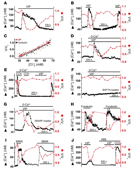
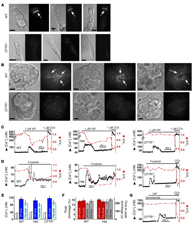
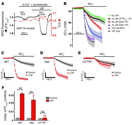
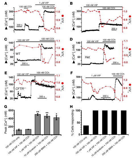
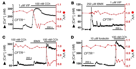
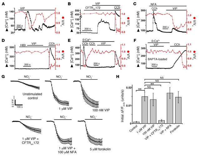
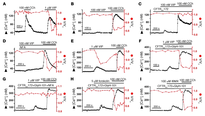
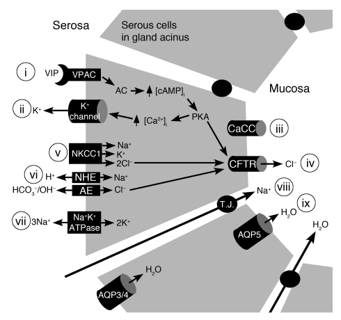
Cystic fibrosis (CF), which is caused by mutations in CFTR, affects many tissues, including the lung. Submucosal gland serous acinar cells are primary sites of fluid secretion and CFTR expression in the lung. Absence of CFTR in these cells may contribute to CF lung pathogenesis by disrupting fluid secretion. Here, we have isolated primary serous acinar cells from wild-type and CFTR-/- pigs and humans without CF to investigate the cellular mechanisms and regulation of fluid secretion by optical imaging. Porcine and human serous cells secrete fluid in response to vasoactive intestinal polypeptide (VIP) and other agents that raise intracellular cAMP levels; here, we have demonstrated that this requires CFTR and a cAMP-dependent rise in intracellular Ca2+ concentration ([Ca2+]i). Importantly, cAMP induced the release of Ca2+ from InsP3-sensitive Ca2+ stores also responsive to cAMP-independent agonists such as cholinergic, histaminergic, and purinergic agonists that stimulate CFTR-independent fluid secretion. This provides two types of synergism that strongly potentiated cAMP-mediated fluid secretion but differed in their CFTR dependencies. First, CFTR-dependent secretion was strongly potentiated by low VIP and carbachol concentrations that individually were unable to stimulate secretion. Second, higher VIP concentrations more strongly potentiated the [Ca2+]i responses, enabling ineffectual levels of cholinergic stimulation to strongly activate CFTR-independent fluid secretion. These results identify important molecular mechanisms of cAMP-dependent secretion, including a requirement for Ca2+ signaling, and suggest new therapeutic approaches to correct defective submucosal gland secretion in CF.


Figures

Figure 1
Figure 1. VIP-stimulated porcine serous cell secretion requires cAMP-dependent Ca2+ signaling.
(A and B) Representative traces showing 1 μM VIP-evoked [Ca2+]i (triangles) and cell volume (circles; normalized to volume at time = 0 [V/Vo]) responses, which were reproducible (B). (C) Cell volume and [Cl]i were linearly correlated during VIP and forskolin exposure. 75 points plotted from 6 forskolin experiments (squares) and 82 points from 5 VIP experiments (circles). SPQ fluorescence changes (Supplemental Figure 1) converted to [Cl]i as described (3, 38). (D) Representative trace showing transient [Ca2+]i elevation and shrinkage during VIP exposure in 0-Ca2+o. (EF) After depletion of Ca2+ stores by repeated stimulation with 100 μM CCh in 0-Ca2+o (E) or in BAPTA-loaded [Ca2+]i-buffered cells (F), VIP exposure in 0-Ca2+ resulted in neither [Ca2+]i elevation nor shrinkage. (G) Cells loaded with SNARF-5F-AM (which does not chelate [Ca2+]i) exhibited normal [Ca2+]i elevation (134 ± 17 nM) and shrinkage (17% ± 2% within 130 ± 22 s; n = 4; NS compared with VIP/0-Ca2+ stimulation as in Figure 1D). (H) Forskolin (10 μM) caused concomitant shrinkage (15% ± 2%; n = 6) and [Ca2+]i elevation (130 ± 11 nM; n = 10; values NS compared with 1 μM VIP). (I) IMBX (250 μM) caused simultaneous [Ca2+]i elevation (97 ± 10 nM; n = 3) and shrinkage (18% ± 2%; values NS compared with 1 μM VIP). (J) VIP-stimulated responses were abolished by H89; responses to 1 μM CCh remained intact.
Figure 2
Figure 2. VIP-activated porcine serous cell secretion requires CFTR.
(AB) Micrographs showing CFTR immunostaining in WT and CFTR–/– porcine tracheal ciliated epithelial cells (A) and serous acini (B). Arrows indicate apical membrane immunofluorescence observed only in WT cells. Scale bars: 5 μm. (C and D) Representative traces showing VIP-evoked (C) and forskolin-evoked (D) volume and [Ca2+]i responses in WT, Het, and CFTR–/– cells. (E) Summary of resting and peak [Ca2+]i. (F) Summary of shrinkage (red) and time to shrinkage (gray) in WT and Het cells. VIP-stimulated shrinkage was 15% ± 1% within 196 ± 15 s (WT; n = 6) and 16% ± 1% within 218 ± 31 s (Het.; n = 6; all values NS). Forskolin-stimulated shrinkage was 16% ± 2% within 228 ± 21 s (WT; n = 14) and 15% ± 2% within 245 ± 18 s (Het.; n = 8; all values NS). (G) Bumetanide (100 μM) did not enhance VIP-evoked shrinkage in CFTR–/– cells.
Figure 3
Figure 3. VIP-induced [Ca2+]i elevation is not required for activation of CFTR-dependent anion permeability in porcine serous cells.
(A) SPQ fluorescence (black inverted triangles) during substitution of Clo with NO3o before and during exposure to 1 μM VIP in presence of 100 μM bumetanide and 0-CO2/HCO3 in 0-Ca2+o, BAPTA-loaded conditions that inhibit shrinkage. (B) Average responses to NO3 substitution (raw traces shown in Supplemental Figure 5) in absence of VIP (n = 7) or after 180–200 s exposure to 1 μM VIP (n = 8) ± 100 μM NFA (n = 5), 10 μM GlyH-101 (n = 5), 30 μM GlyH-101 (n = 4), or 10 μM CFTRinh172 (n = 9). Fluorescence normalized to that at time = 0 (F/F(t=0)) and converted to [Cl]i (4) before averaging. (CE) Cl permeability measured as above in WT (C), Het (D), or CFTR–/– (E) tracheal cells either unstimulated or stimulated for 180–200 s with 1 μM VIP. SPQ fluorescence plotted inversely (downward deflection = increase in SPQ fluorescence = decrease in [Cl]i). (F) Initial rates of SPQ fluorescence increase in unstimulated (control) and 1 μM VIP-stimulated WT (n = 12), Het (n = 7), and CFTR–/– (n = 8) cells. Red asterisks indicate significance of VIP-stimulated rates (red bars) compared with WT (Dunnett’s test). Black asterisks indicate significance between control and VIP-stimulated rates (gray versus red bar; Student’s t test) within each genotype. *P < 0.05; **P < 0.01.
Figure 4
Figure 4. VIP potentiates low [CCh]-evoked responses in porcine serous cells.
(A) In 3 of 8 bronchial cells, 100 nM CCh elicited [Ca2+]i responses similar in magnitude to those evoked by 1 μM VIP; however, only VIP elicited shrinkage. (B) 100 nM VIP elicited smaller [Ca2+]i elevations but no shrinkage. Subsequent 100 nM CCh elicited Ca2+-rise and shrinkage in 100% of cells. (CE) Neonatal tracheal cells stimulated with 100 nM VIP plus 100 nM CCh exhibited [Ca2+]i elevations and CFTR-dependent shrinkage. Resting [Ca2+]i was 34 ± 2 nM (WT; n = 13), 30 ± 2 nM (Het; n = 12), and 30 ± 6 nM (CFTR–/–; n = 7). 100 nM VIP caused [Ca2+]i elevation of 50 ± 4 nM (WT), 45 ± 3 nM (Het), and 56 ± 16 nM (CFTR–/–). 100 nM CCh (in continued presence of VIP) elicited peak [Ca2+]i responses of 128 ± 6 nM (WT), 132 ± 77 nM (Het), and 130 ± 16 nM (CFTR–/–). Plateau [Ca2+]i was 97 ± 5 nM (WT), 96 ± 6 nM (Het), and 83 ± 8 nM (CFTR–/–). WT and Het cells shrank; CFTR–/– cells did not. (F) After 1 μM VIP, 100 nM CCh elicited higher [Ca2+]i peak (492 ± 24 nM) and enhanced shrinkage in all cells. (G and H). [Ca2+]i responses during 100 nM CCh stimulation of WT bronchial serous cells ± cAMP agonists. Asterisks represent significance compared with 100 nM CCh alone (Dunnett’s test, *P < 0.05; **P < 0.01).
Figure 5
Figure 5. Strong cAMP stimulation restores low [CCh]-evoked secretion in CFTR–/– porcine tracheal gland serous cells.
(A) In CFTR–/– cells, high [VIP] (1 μM) elicited exaggerated 100 nM CCh-stimulated [Ca2+]i rise (340 ± 42 nM) correlated with shrinkage (19% ± 2%). (B and C) IBMX caused [Ca2+]i elevation (104 ± 18 nM) but not shrinkage in CFTR–/– cells (n = 4). No additive effects were observed with VIP (B), but 100 nM CCh-evoked [Ca2+]i responses were potentiated (303 ± 13 nM; n = 4; C), causing CFTR–/– cells to shrink (20% ± 2%). (D) Similar results observed with 10 μM forskolin pretreatment. Subsequent 100 nM CCh evoked-peak [Ca2+]i response (455 ± 30 nM) was correlated with shrinkage (22% ± 2%; n = 5).
Figure 6
Figure 6. VIP stimulation of human nasal gland serous cell secretion requires both PKA-dependent Ca2+ signaling and Ca2+-independent CFTR activation.
(A) VIP (1 μM) caused cell shrinkage that was temporally correlated with elevation of [Ca2+]i. (B and C) VIP-activated shrinkage was completely blocked by CFTRinh172 (12 μM, B); CCh-activated shrinkage was inhibited by NFA (150 μM, C). (D) VIP-activated [Ca2+]i elevation and shrinkage were both abolished by H89 (10 μM). (E) After intracellular Ca2+ store depletion by stimulation with 100 μM CCh in 0-Ca2+o, subsequent stimulation with 1 μM VIP in 0-Ca2+o elicited neither [Ca2+]i nor shrinkage responses. Reintroduction of Ca2+o caused a transient [Ca2+]i elevation and shrinkage. (F) BAPTA-loading inhibited [Ca2+]i elevation and shrinkage during stimulation with VIP in 0-Ca2+o. Reintroduction of Ca2+o in the presence of CCh caused a gradual rise of [Ca2+]i, activating shrinkage. (G) NO3 substitution was performed in BAPTA- and SPQ-loaded cells under 0-Ca2+o/0-HCO3o/100 μM bumetanide conditions (identical to Figure 3). VIP (100 nM and 1 μM) activated an approximately 15-fold increase in anion permeability that was mimicked by 5 μM forskolin and inhibited by 12 μM CFTRinh172 but not by 150 μM NFA. Average traces shown (plotted inversely: downward deflection = increase in fluorescence = decrease in [Cl]i) (H) Summary of initial rates of SPQ ΔF/F(t=0) (units × s–1) upon introduction of NO3. **P < 0.01.
Figure 7
Figure 7. Human nasal gland serous acinar cells exhibit dose-dependent synergy between VIP and low-level cholinergic stimulation.
(A) Stimulation with either 100 nM CCh or 1 μM VIP caused comparable [Ca2+]i elevations in human serous cells, but only VIP caused cell shrinkage. (B) While a lower [VIP] (100 nM) had minimal effect on [Ca2+]i and no effect on volume, it synergistically activated shrinkage in response to 100 nM CCh despite having no significant effect on the [Ca2+]i response. (C and D) Shrinkage in response to 100 nm VIP + 100 nM CCh was inhibited by 12 μM CFTRinh172 (C) but not by 150 μM NFA (D). (EG). Higher [VIP] (1 μM) markedly potentiated the [Ca2+]i elevation in response to 100 nM CCh (E), activating shrinkage that was insensitive to the presence of 12 μM CFTRinh172 plus 5 μM GlyH-101 (F) but was blocked by NFA (G). (H and I) Strong cAMP stimulation with 5 μM forskolin (H) or 100 μM IBMX (I) likewise potentiated 100 nM CCh-evoked [Ca2+]i responses, activating CFTR-independent shrinkage.
Figure 8
Figure 8. Model of VIP/cAMP evoked fluid secretion in porcine and human airway gland serous acinar cells.
Binding of VIP to VPAC receptors (i) activates adenylyl cyclase–mediated (AC-mediated) elevation of [cAMP]i, causing PKA-stimulated elevation of [Ca2+]i required for activation of basolateral K+ channels (ii). This [Ca2+]i response is insufficient to activate CaCCs (iii). Thus, Cl secretion requires PKA-dependent activation of CFTR (iv). As during cholinergic stimulation, transepithelial Cl secretion is sustained by NKCC1 (v) and NHE/AE (vi), expressed on the basolateral membrane (4) and driven by Na+ gradient established by the Na+/K+ ATPase (vii). While AE function was not elucidated directly in this study, canonical models of epithelial Cl secretion (reviewed in ref. 44) dictate that NHE-mediated alkalinization drives Cl/HCO3 exchange resulting in Cl uptake to sustain secretion. Secretion of Cl drives movement of Na+ through a paracellular (tight junction; T.J.) pathway (viii) drawing osmotically obliged water into the gland lumen (ix) paracellularly or transcellularly through aquaporins (AQP; localization based on ref. 45). Our data suggest that activation of CaCC(s), either directly or indirectly through agents that enhance cAMP-activated [Ca2+]i signals, could restore fluid secretion in serous cells lacking functional CFTR.

Comment in

References

    1. Ballard ST, Spadafora D. Fluid secretion by submucosal glands of the tracheobronchial airways. Respir Physiol Neurobiol. 2007;159(3):271–277. doi: 10.1016/j.resp.2007.06.017. - DOI - PMC - PubMed
    1. Engelhardt JF, et al. Submucosal glands are the predominant site of CFTR expression in the human bronchus. Nat Genet. 1992;2(3):240–248. doi: 10.1038/ng1192-240. - DOI - PubMed
    1. Lee RJ, Limberis MP, Hennessy MF, Wilson JM, Foskett JK. Optical imaging of Ca2+-evoked fluid secretion by murine nasal submucosal gland serous acinar cells. . J Physiol. 2007;582(pt 3):1099–1124. doi: 10.1113/jphysiol.2007.131995. - DOI - PMC - PubMed
    1. Lee RJ, Foskett JK. Mechanisms of Ca2+-stimulated fluid secretion by porcine bronchial submucosal gland serous acinar cells. . Am J Physiol Lung Cell Mol Physiol. 2010;298(2):L210–L231. doi: 10.1152/ajplung.00342.2009. - DOI - PubMed
    1. Joo NS, Irokawa T, Robbins RC, Wine JJ. Hyposecretion, not hyperabsorption, is the basic defect of cystic fibrosis airway glands. J Biol Chem. 2006;281(11):7392–7398. doi: 10.1074/jbc.M512766200. - DOI - PubMed

Publication types

MeSH terms

Substances