. 2010 Sep;120(9):3389-400.
doi: 10.1172/JCI43350. Epub 2010 Aug 25.

The poly(A)-binding protein partner Paip2a controls translation during late spermiogenesis in mice

Affiliations

The poly(A)-binding protein partner Paip2a controls translation during late spermiogenesis in mice

Akiko Yanagiya et al. J Clin Invest. 2010 Sep.

Abstract

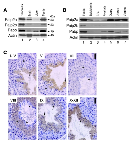
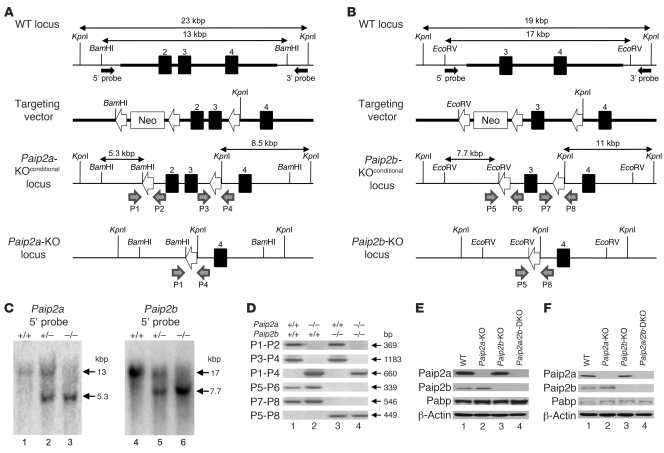
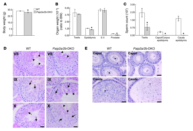
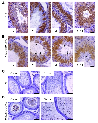
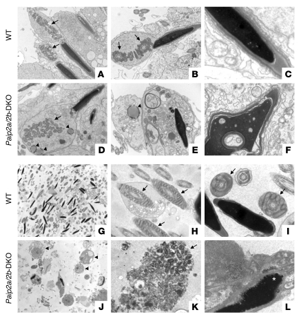
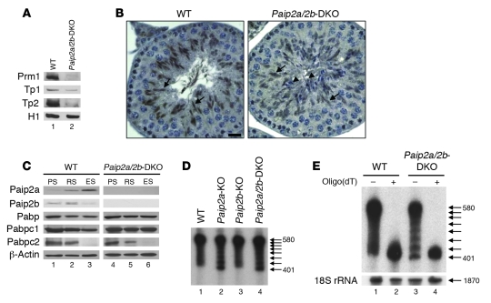
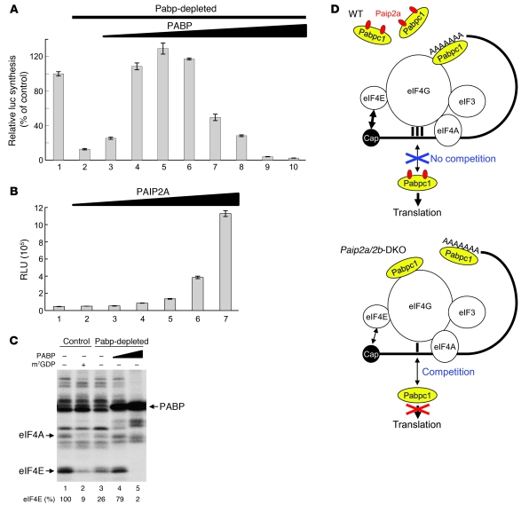
Translational control plays a key role in late spermiogenesis. A number of mRNAs encoding proteins required for late spermiogenesis are expressed in early spermatids but are stored as translationally inactive messenger ribonucleoprotein particles (mRNPs). The translation of these mRNAs is associated with shortening of their poly(A) tail in late spermiogenesis. Poly(A)-binding protein (Pabp) plays an important role in mRNA stabilization and translation. Three Pabp-interacting proteins, Paip1, Paip2a, and Paip2b, have been described. Paip2a is expressed in late spermatids. To investigate the role of Paip2 in spermiogenesis, we generated mice with knockout of either Paip2a or Paip2b and double-KO (DKO) mice lacking both Paip2a and Paip2b. Paip2a-KO and Paip2a/Paip2b-DKO mice exhibited male infertility. Translation of several mRNAs encoding proteins essential to male germ cell development was inhibited in late spermiogenesis in Paip2a/Paip2b-DKO mice, resulting in defective elongated spermatids. Inhibition of translation in Paip2a/Paip2b-DKO mice was caused by aberrant increased expression of Pabp, which impaired the interaction between eukaryotic initiation factor 4E (eIF4E) and the cap structure at the 5' end of the mRNA. We therefore propose a model whereby efficient mRNA translation in late spermiogenesis occurs at an optimal concentration of Pabp, a condition not fulfilled in Paip2a/Paip2b-DKO mice.


Figures

Figure 1
Figure 1. Paip2a and Paip2b expression in mouse.
(A) Tissue distribution of Paip2a, Paip2b, and Pabp in pancreas, brain, liver, and testis of WT mouse by Western blot analysis. (B) Paip2a, Paip2b, and Pabp expression in reproductive tissues of WT mouse. S.V., seminal vesicle. (C) Paip2 expression in WT testis visualized by immunohistochemistry. Asterisks show RSs. Arrows indicate elongated spermatids, and arrowheads show mature sperm. Scale bars: 25 μm. Paip2a-specific and Paip2b-specific antibodies were used for Western blotting. Affinity-purified Paip2 antibody that mainly reacts with Paip2a and weakly cross-reacts with Paip2b was used for immunohistochemistry.
Figure 2
Figure 2. Generation of Paip2a/Paip2b-KO mouse.
(A) Targeted disruption of Paip2a. Exons 2, 3, and 4 are depicted as filled boxes. The targeting regions are shown as bold lines. Filled arrows indicate 5′ probe and 3′ probe for Southern blot analysis. Open arrows indicate LoxP sites. Gray arrows indicate primers for genotyping (P1, P2, P3, and P4). (B) Targeted disruption of Paip2b. Exons 3 and 4 are shown as filled boxes. Gray arrows indicate primers for genotyping (P5, P6, P7, and P8). (C) Southern blot analysis of WT, Paip2a heterozygote, Paip2a-KO, Paip2b heterozygote, and Paip2b-KO mice probed by 5′ probes (see also Supplemental Figure 1). (D) Genotyping of WT, Paip2a-KO, Paip2b-KO, and Paip2a/Paip2b-DKO mice. Primer sets of P1 and P2, P3 and P4, and P1 and P4 were used to detect the first and second LoxP sites and the recombined loxP site of Paip2a, respectively. Primer sets of P5 and P6, P7 and P8, and P5 and P8 were used to detect the first LoxP site, the second LoxP site, and the recombined loxP site of Paip2b, respectively. (E) Western blot analysis of Paip2a, Paip2b, and Pabp in testes. (F) Western blot analysis of Paip2a, Paip2b, and Pabp in epididymides.
Figure 3
Figure 3. Phenotypes of Paip2a/Paip2b-KO mice.
(A) Body weights of WT and Paip2a/Paip2b-DKO mice. Values are mean ± SEM of 20–23 mice at 10–12 weeks old. *P < 0.05 using an unpaired t test. (B) Reproductive organ weights of WT and Paip2a/Paip2b-DKO mice. Organ weights per body weight (BW) are shown. Values are mean ± SEM of 10–16 mice for testis and epididymis and 8–14 mice for seminal vesicle and prostate. *P < 0.05 using an unpaired t test. (C) Sperm count in testes, caput/corpus epididymides, and cauda epididymides of WT and Paip2a/Paip2b-DKO mice. Values are mean ± SEM of 5 mice. *P < 0.05 using an unpaired t test. (D and E) Histology of testes (D) and caput and cauda epididymides (E) from WT and Paip2a/Paip2b-DKO mice by H&E staining. Arrows indicate elongating spermatids; arrowheads in WT mice indicate mature sperm released from the epithelium at stage VII; and arrowheads in Paip2a/Paip2b-DKO mice show sperm that could not be released into the lumen in later stages (D). The asterisks indicate mature sperm in the lumen of WT epididymis (E). Scale bars: 25 μm.
Figure 4
Figure 4. Abnormal Pabp expression in late spermiogenesis in Paip2a/Paip2b-DKO mice.
(A and B) Immunohistochemistry of Pabp in testes of WT (A) and Paip2a/Paip2b-DKO (B) mice. Stages I–IV, V, VII, and X–XII are shown. Arrows show aberrant Pabp expression in elongated spermatids at stages V and VII of Paip2a/Paip2b-DKO mice. An asterisk indicates a layer of aberrant elongated spermatids in which Pabp is slightly expressed at stages X–XII in Paip2a/Paip2b-DKO testis. Scale bars: 25 μm. (C and D) Immunohistochemistry of Pabp in caput and cauda epididymides of WT (C) and Paip2a/Paip2b-DKO (D) mice. Scale bars: 50 μm.
Figure 5
Figure 5. Abnormal chromatin condensation and mitochondrial alignment in Paip2a/Paip2b-DKO mice.
(AF) Spermatids in testes from WT (AC) and Paip2a/Paip2b-DKO (DF) mice at stage VII of spermatogenesis. Mitochondria (arrows) and aberrant spherical structures in Paip2a/Paip2b-DKO mice (arrowheads) are shown. (GL) Spermatozoa in cauda epididymides from WT (GI) and Paip2a/Paip2b-DKO (JL) mice. Mitochondria (arrows), ectopic residual bodies (arrowheads), and bubble-like structures in nucleus (asterisk) are shown. Original magnification, A and D: ×8100, B and E: ×10,200, C and F: ×24,400, G and J: ×2,120, H and K: ×13,800, I and L: ×24,400.
Figure 6
Figure 6. Translation inhibition in late spermiogenesis of Paip2a/Paip2b-DKO mice.
(A) Levels of basic nuclear proteins (Prm1, Tp1, Tp2, and histone H1) from testes of WT and Paip2a/Paip2b-DKO mice were analyzed by acid-urea polyacrylamide gel electrophoresis. (B) Tp2 expression in testes at stages X–XII from WT and Paip2a/Paip2b-DKO mice analyzed by immunohistochemistry. Arrows indicate Tp2-positive spermatids, and arrowheads indicate Tp2-negative spermatids. Scale bar: 10 μm. (C) Expression of Paip2a, Paip2b, Pabpc1, and Pabpc2 in PSs, RSs, and ESs of WT (lanes 1–3) and Paip2a/Paip2b-DKO (lanes 4–6) mice analyzed by Western blotting. (D) Prm1 mRNA in testes from WT, Paip2a-KO, Paip2b-KO, and Paip2a/Paip2b-DKO mice analyzed by Northern blot analysis. Numbers on the right denote nucleotides, and arrows indicate ladder RNA fragments. (E) Ladder RNA fragments were generated by cleavage of poly(A) tail. Total testis RNA of WT and Paip2a/Paip2b-DKO mice was incubated without (–) or with (+) oligo(dT) and then treated with RNase H, followed by Northern blot analysis. Ribosomal 18S RNA (1,870 nucleotides) was used as a control for the absence of nonspecific RNA degradation during this experiment.
Figure 7
Figure 7. Translational control of late spermiogenesis by Paip2a.
(A) Excess amounts of PABP inhibit translation in vitro. Control (lane 1) or Pabp-depleted Krebs-2 cell extracts (lanes 2–10) were programmed with capped Luc(A98) mRNA (2 μg/ml). The translation in Pabp-depleted Krebs-2 extract was performed in the presence of increasing concentrations of recombinant PABP (lanes 2–10; 0, 1.5, 3.0, 4.5, 6.0, 12, 18, 24, and 30 μg/ml). Relative light units were measured, and the value in control (1,034,812 RLU) was set as 100%. Data are presented as mean ± SD. (B) Translation of capped Luc(A98) mRNA in Pabp-depleted Krebs-2 extract supplemented with excess PABP (30 μg/ml) was performed in the presence of increasing concentrations of recombinant GST-PAIP2A (lanes 1–7; 0, 2.5, 5.0, 10, 15, 20, and 30 μg/ml). Data are presented in RLU as mean ± SD. (C) Crosslinking of initiation factors to the cap structure. Control RRL (lanes 1 and 2) or Pabp-depleted RRL (lanes 3–5) was preincubated with control buffer (–) (lanes 1 and 3), 0.6 mM m7GDP (+) (lane 2), and PABP (15 or 30 μg/ml) (lanes 4 and 5, respectively). Oxidized [32P]cap-labeled Luc(A+) mRNA was then added. After reduction with NaIO4 and nuclease digestion, labeled proteins were analyzed by SDS-PAGE and autoradiography. Relative efficiencies of eIF4E crosslinking to the cap structure are indicated at the bottom. The value in control RRL (lane 1) was set as 100%. (D) Model of molecular mechanism of translational control in late spermiogenesis by Paip2a.

Comment in

  • Too much PABP, too little translation.
    Kini HK, Vishnu MR, Liebhaber SA. Kini HK, et al. J Clin Invest. 2010 Sep;120(9):3090-3. doi: 10.1172/JCI44091. Epub 2010 Aug 25. J Clin Invest. 2010. PMID: 20739750 Free PMC article.

Similar articles

Cited by

References

    1. Kerr JB, Loveland KL, O’Bryan MK, de Kretser DM. Cytology of the testis and intrinsic control mechanisms. In: Neill JD, ed.Physiology of Reproduction . 3rd ed. Maryland Heights, Missouri, USA: Elsevier Press; 2006:1.
    1. Hecht NB. Molecular mechanisms of male germ cell differentiation. Bioessays. 1998;20(7):555–561. doi: 10.1002/(SICI)1521-1878(199807)20:7<555::AID-BIES6>3.0.CO;2-J. - DOI - PubMed
    1. Steger K. Transcriptional and translational regulation of gene expression in haploid spermatids. Anat Embryol (Berl). 1999;199(6):471–487. - PubMed
    1. Kleene KC. Multiple controls over the efficiency of translation of the mRNAs encoding transition proteins, protamines, and the mitochondrial capsule selenoprotein in late spermatids in mice. Dev Biol. 1993;159(2):720–731. doi: 10.1006/dbio.1993.1277. - DOI - PubMed
    1. Penttila TL, Yuan L, Mali P, Hoog C, Parvinen M. Haploid gene expression: temporal onset and storage patterns of 13 novel transcripts during rat and mouse spermiogenesis. Biol Reprod. 1995;53(3):499–510. doi: 10.1095/biolreprod53.3.499. - DOI - PubMed

Publication types