. 2010 Sep 16;467(7313):347-51.
doi: 10.1038/nature09323. Epub 2010 Aug 25.

The structure of (CENP-A-H4)(2) reveals physical features that mark centromeres

Affiliations

The structure of (CENP-A-H4)(2) reveals physical features that mark centromeres

Nikolina Sekulic et al. Nature. .

Abstract

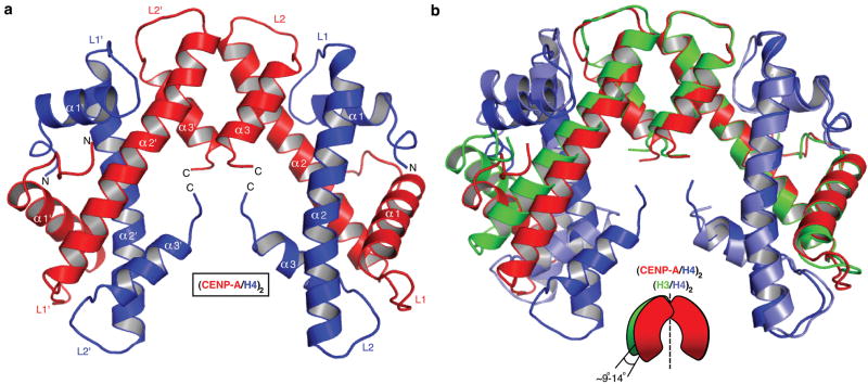
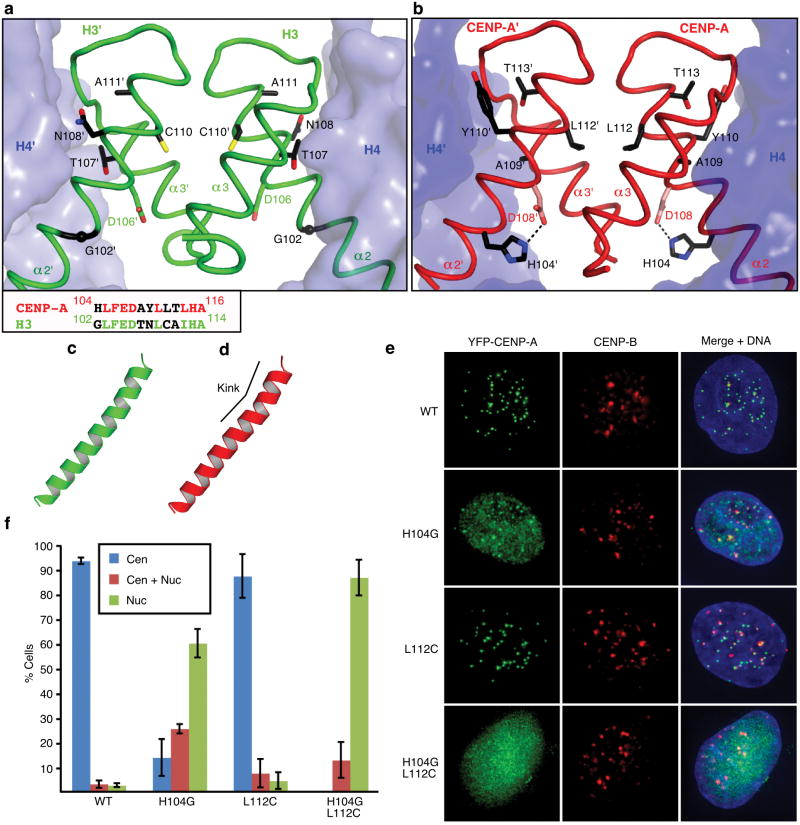
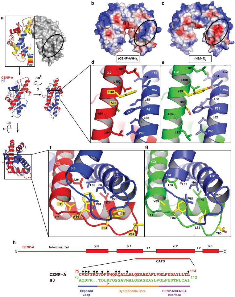
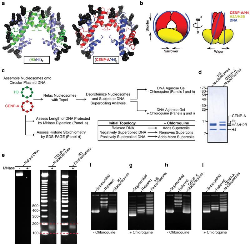
Centromeres are specified epigenetically, and the histone H3 variant CENP-A is assembled into the chromatin of all active centromeres. Divergence from H3 raises the possibility that CENP-A generates unique chromatin features to mark physically centromere location. Here we report the crystal structure of a subnucleosomal heterotetramer, human (CENP-A-H4)(2), that reveals three distinguishing properties encoded by the residues that comprise the CENP-A targeting domain (CATD; ref. 2): (1) a CENP-A-CENP-A interface that is substantially rotated relative to the H3-H3 interface; (2) a protruding loop L1 of the opposite charge as that on H3; and (3) strong hydrophobic contacts that rigidify the CENP-A-H4 interface. Residues involved in the CENP-A-CENP-A rotation are required for efficient incorporation into centromeric chromatin, indicating specificity for an unconventional nucleosome shape. DNA topological analysis indicates that CENP-A-containing nucleosomes are octameric with conventional left-handed DNA wrapping, in contrast to other recent proposals. Our results indicate that CENP-A marks centromere location by restructuring the nucleosome from within its folded histone core.


Figures

Figure 1
Figure 1. Crystal structure of the (CENP-A/H4)2 heterotetramer
a, Ribbon diagram of the (CENP-A/H4)2 heterotetramer. The structure has clear electron density for residues 59-134 of CENP-A and 24-92 of H4. b, Overlay of (CENP-A/H4)2 with (H3/H4)2 (PDB ID 1KX515) was done with a secondary structure mapping (SSM) algorithm performed with one CENP-A molecule and one H3 molecule from each complex. CENP-A and H3 molecules on the right are overlaid. Similar results are obtained if one molecule of H4 from each complex is used for SSM (rmsd=~0.9 Å in both cases).
Figure 2
Figure 2. The residues involved in the rotated CENP-A/CENP-A interface are essential for centromere targeting
Detail of H3/H3, a, and CENP-A/CENP-A, b, interfaces with side chains of 5 non-conserved residues in α2 helix at the interface. An alignment of residues in the C-terminal portion of the α2 helix are shown in the box at lower left in panel a. c and d, the α2 helices of H3, c, and CENP-A, d. e, Centromere targeting of WT and mutant versions of CENP-A. f, Quantitation of targeting experiment. For each version of CENP-A, 3 or more experiments were conducted in which a total of >250 cells were analyzed for localization (Cen = exclusively centromeric; Cen + Nuc = both centromere and nucleoplasm staining; Nuc = nucleoplasm only [i.e. no enrichment at centromeres]). Values are plotted +/- s.d.
Figure 3
Figure 3. Surface and internal structural features unique to CENP-A-containing complexes
a, The rates of H/DX remapped from the original data are plotted onto the crystal structure of the (CENP-A/H4)2 heterotetramer (left dimer half is shown as ribbon and right dimer half is in surface representation). b and c, Calculated electrostatic potential on the surface of (CENP-A/H4)2, b, and (H3/H4)2, c. The region corresponding to the positively charged bulge in the L1 of CENP-A is circled in each structure. d and e, Hydrophobic interactions between the α2 helices of CENP-A (red) and H4 (blue), d, and α2 helices of H3 (green) and H4 (light blue) e. f and g, Hydrophobic interactions at the helical bundle surrounding the interface of L1 of CENP-A and L2 of H4, f, and L1 of H3 and L2 of H4, g. h, Diagram of the structural differences between CENP-A and H3. Black circles indicate surface exposed residues. The yellow star highlights Arg83 from H3 that inserts into the minor groove of nucleosomal DNA.
Figure 4
Figure 4. The (CENP-A/H4)2 heterotetramer assembles with H2A/H2B dimers into an octameric nucleosome with conventional handedness of DNA wrapping
a, Basic residues that form contacts with nucleosomal DNA are highlighted on (H3/H4)2 (left) and the putative DNA binding surface of (CENP-A/H4)2 (right). b, Model highlighting alterations in nucleosome structure based on the structure of (CENP-A/H4)2. c, Scheme for nucleosome assembly and analysis. d, Histone content of assembled H3- and CENP-A-containing nucleosomes. e, Digestion of nucleosome arrays with micrococcal nuclease reveals that both H3- and CENP-A-containing nucleosomes protect ~150 bp of DNA. f-i, Topological analysis of H3- containing (f and g) and CENP-A-containing (h and i) nucleosomes. Analysis by gel electrophoresis in the absence (f and h) or presence (g and i) of chloroquine reveals that both H3-containing and CENP-A-containing nucleosomes wrap DNA in a left-handed manner.

Similar articles

Cited by

References

    1. Black BE, Bassett EA. The histone variant CENP-A and centromere specification. Curr Opin Cell Biol. 2008;20:91–100. - PubMed
    1. Black BE, et al. Structural determinants for generating centromeric chromatin. Nature. 2004;430:578–582. - PubMed
    1. Mizuguchi G, Xiao H, Wisniewski J, Smith MM, Wu C. Nonhistone Scm3 and histones CenH3-H4 assemble the core of centromere-specific nucleosomes. Cell. 2007;129:1153–1164. - PubMed
    1. Williams JS, Hayashi T, Yanagida M, Russell P. Fission yeast Scm3 mediates stable assembly of Cnp1/CENP-A into centromeric chromatin. Mol Cell. 2009;33:287–298. - PMC - PubMed
    1. Dalal Y, Wang H, Lindsay S, Henikoff S. Tetrameric structure of centromeric nucleosomes in interphase Drosophila cells. PLoS Biol. 2007;5:e218. - PMC - PubMed

Publication types

MeSH terms