. 2010 Aug 26;466(7310):1069-75.
doi: 10.1038/nature09320.

Ataxin-2 intermediate-length polyglutamine expansions are associated with increased risk for ALS

Affiliations

Ataxin-2 intermediate-length polyglutamine expansions are associated with increased risk for ALS

Andrew C Elden et al. Nature. .

Abstract

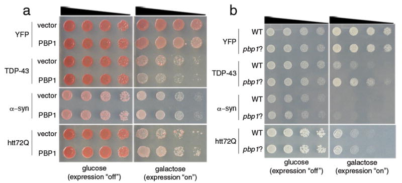
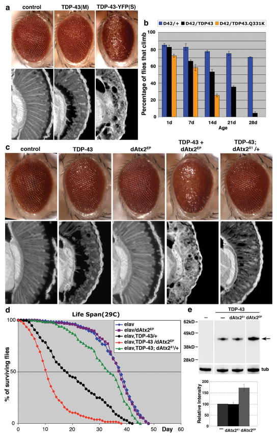
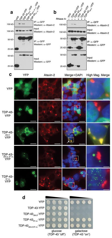
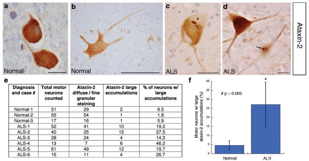
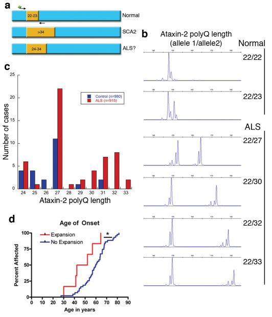
The causes of amyotrophic lateral sclerosis (ALS), a devastating human neurodegenerative disease, are poorly understood, although the protein TDP-43 has been suggested to have a critical role in disease pathogenesis. Here we show that ataxin 2 (ATXN2), a polyglutamine (polyQ) protein mutated in spinocerebellar ataxia type 2, is a potent modifier of TDP-43 toxicity in animal and cellular models. ATXN2 and TDP-43 associate in a complex that depends on RNA. In spinal cord neurons of ALS patients, ATXN2 is abnormally localized; likewise, TDP-43 shows mislocalization in spinocerebellar ataxia type 2. To assess the involvement of ATXN2 in ALS, we analysed the length of the polyQ repeat in the ATXN2 gene in 915 ALS patients. We found that intermediate-length polyQ expansions (27-33 glutamines) in ATXN2 were significantly associated with ALS. These data establish ATXN2 as a relatively common ALS susceptibility gene. Furthermore, these findings indicate that the TDP-43-ATXN2 interaction may be a promising target for therapeutic intervention in ALS and other TDP-43 proteinopathies.


Figures

Figure 1
Figure 1
Pbp1 is a dose-sensitive modifier of TDP-43 toxicity in yeast. a) Spotting assays with yeast TDP-43 showing toxicity. Five-fold serial dilutions of yeast cells spotted onto glucose (expression repressed) or galactose (expression induced). Upregulation of PBP1 enhances TDP-43 toxicity. Whereas PBP1 has no effect on yeast viability when expressed with the control protein YFP, when co-expressed with TDP-43, it enhances toxicity. Enhancement is specific because PBP1 does not affect the toxicity of a pathogenic Huntington fragment (htt72Q) or α–synuclein. b) Spotting assays with yeast TDP-43 showing that PBP1 deletion (pbp1Δ) suppresses TDP-43 toxicity. Whereas expression of TDP-43 from a plasmid in WT yeast was toxic, this was mitigated in pbp1Δ cells. The effect was specific because α–syn or htt72Q toxicity was not suppressed by pbp1Δ
Figure 2
Figure 2
Ataxin-2 is a dose-sensitive modifier of TDP-43 toxicity in Drosophila. a) Expression of TDP-43 caused a dose-dependent disruption of retinal structure. Genotypes: gmr-GAL4(YH3) in trans to UAS-YFP (control), UAS-TDP-43(M) or UAS-TDP-43-YFP(S). (M) and (S) are moderate and strong TDP-43 expression, respectively (see Methods and Fig. S2). b) TDP-43 caused motility deficits when expressed in motor neurons. TDP-43.Q331K causes a more severe loss of motility than the WT protein at the same level of expression (see Fig. S2). Genotypes: D42-GAL4 in trans to +, UAS-TDP-43 or UAS-TDP-43.Q331K. c) dAtx2 modulates TDP-43 toxicity. Flies expressing TDP-43 or dAtx2 alone (dAtx2EP) have a mild effect on retinal structure. TDP-43 toxicity is more severe upon upregulation of dAtx2 (dAtx2EP3145). TDP-43 toxicity is markedly mitigated upon reduction of dAtx2 (flies in trans to null allele dAtx2X1). Genotypes: control: gmr-GAL4/UAS-YFP. TDP-43: UAS-TDP-43(M)/+, gmr-GAL4(YH3)/+. dAtx2EP: gmr-GAL4(YH3)/dAtx2EP3145. TDP43+dAtx2EP: UAS-TDP-43(M)/+, gmr-GAL4(YH3)/dAtx2EP3145. TDP-43;dAtx2X1/+: UAS-TDP-43(M)/+, gmr-GAL4(YH3)/dAtx2X1. dAtx2X1/+ has wild type retinal structure. d) dAtx2 modulates reduced lifespan conferred by TDP-43. Expression of TDP-43 in the nervous system reduces lifespan (black, compared to normal in blue). Upregulation of dAtx2 causes more rapid death (red, compared to TDP-43 in black). Upregulation of dAtx2 on its own has no effect (purple). Reduction of dAtx2 significantly extends lifespan (green, compared to TDP-43 in black). Heterozygous loss of dAtx2 on its own has no effect (not shown). quantitative-RT-PCR showed that TDP-43 expression has no effect on levels of dAtx2 transcript in dAtx2EP flies. Genotypes. elav/+: elavC155/Y. elav/dAtx2EP: elavC155/Y; +/dAtx2EP3145. elav, TDP-43/+: elavC155/Y; UAS-TDP-43(M)/+. elav, TDP-43/dAtx2EP: elavC155/Y; UAS-TDP-43(M)/+; +/dAtx2EP3145. elav, TDP-43; dAtx2X1/+: elavC155/Y; UAS-TDP-43(M)/+; +/dAtx2X1. e) TDP-43 immunoblot upon dAtx2 modulation. Reduction of dAtx2 has no effect on TDP-43 levels, whereas upregulation of dAtx2 enhances TDP-43 protein levels. dAtx2 has no effect on the transgene expression system and 2x TDP-43 does not produce the same phenotype as TDP-43+dAtx2EP (see Figs. S2 and S3). Genotypes: —, control lane. TDP-43 lanes, —: UAS-TDP-43(M)/+, gmr-GAL4(YH3)/+. dAtx2X1:UAS-TDP-43(M)/+, gmr-GAL4(YH3)/dAtx2X1. dAtx2EP: UAS-TDP-43(M)/+, gmr-GAL4(YH3)/dAtx2EP3145. Bottom, quantitation of immunoblots from independent experiments; normalized to tubulin.
Figure 3
Figure 3
Ataxin-2 and TDP-43 interact in a manner dependent on RNA. a) TDP-43 and Ataxin-2 associate in mammalian cells in a manner dependent on the RRMs. HEK293T cells were transfected with plasmids encoding YFP, TDP-43-YFP, TDP-43 ΔNLS-YFP (NLS mutant localizes to cytoplasm), TDP-43 ΔNLS,5F→L-YFP (NLS mutant + RNA-binding mutant), or TDP-435F→L-YFP (RNA-binding mutant). Protein was immunoprecipitated with anti-GFP antibody, and then subjected to immunoblotting with anti-Ataxin-2 to detect endogenous Ataxin-2. Whereas TDP-43 and TDP-43 ΔNLS both interact with Ataxin-2, RNA-binding mutant versions do not. b) Co-IP in HEK293T cells as in (a), but now with lysates treated with RNase. The interaction between Ataxin-2 and TDP-43 seen normally was abolished upon RNase treatment. c) HEK293T cells transfected with YFP-tagged WT and mutant TDP-43 constructs then immunostained for endogenous Ataxin-2. Normally, Ataxin-2 is localized to the cytoplasm forming occasional cytoplasmic accumulations. TDP-43 localized to the nucleus in a diffuse pattern. TDP-43 ΔNLS localized to the cytoplasm where it occasionally formed cytoplasmic aggregates; these aggregates always co-localized with Ataxin-2 cytoplasmic accumulations (arrow). Abolishing the ability of TDP-43 to interact with RNA (TDP-43 ΔNLS, 5F→L or TDP-435F→L) eliminated Ataxin-2 colocalization (arrowheads). TDP-435F→L-YFP was restricted to the nucleus where it formed multiple foci. Scale bar is 2.5 μm for merge panels and 0.5 μm for high mag. merge panels. d) Yeast spotting assays for TDP-43 toxicity. Whereas WT and TDP-43 ΔNLS constructs are toxic, mutations of TDP-43 that prevent RNA binding abolish toxicity.
Figure 4
Figure 4
Ataxin-2 localization is perturbed in ALS patient neurons. a–d, immunostaining for Ataxin-2 in spinal cord. a–b) In control spinal cord neurons, Ataxin-2 is localized throughout the cytoplasm in a diffuse pattern (c–f and Fig. S10). In ALS spinal cord neurons, Ataxin-2 was present in distinct cytoplasmic accumulations (arrows). In some cases, Ataxin-2-positive accumulations were adjacent to clearings indicative of TDP-43 aggregates (* in (b)). e, f) Quantitation of Ataxin-2 large accumulations in control (normal) vs. ALS spinal cord neurons. 27.2 +/− 12.3% of spinal cord neurons in ALS patients had large accumulations of Ataxin-2 compared to 4.7+/− 2.6% of control neurons. ALS patients with normal (ALS 1–3) and intermediate-length (ALS 4–6) Ataxin-2 polyQ repeats (See Fig. 5) were included and the Ataxin-2 pathology was not significantly different. An ALS case with both an SOD1 mutation and Ataxin-2 polyQ expansion (27Q) was included (case ALS-4). Scale bar, 1.25 μm for a; 5 μm for b–d.
Figure 5
Figure 5
Intermediate-length Ataxin-2 polyQ expansions linked to ALS. a) The ATXN2 gene contains a trinucleotide repeat encoding polyQ. The repeat length is normally 22–23Q. Expansions of > 34 cause SCA2. We hypothesized that intermediate-length polyQ expansions (e.g. 24–34) could be linked to ALS. The Ataxin-2 polyQ length was defined by Genescan analysis of ALS cases and neurologically normal controls (for details, see Table 1 and Methods). b) Representative examples of Genescan analysis of polyQ lengths from control and ALS cases. c) The distribution of Ataxin-2 polyQ repeat lengths in ALS and control cases. PolyQ lengths ≥27 are significantly enriched in ALS vs. controls. Additionally, polyQ lengths >31 were never observed in our controls but we found 10 ALS patients above this threshold. d) In a selected cohort of ALS patients (n=65), those with Ataxin-2 polyQ expansions showed significantly lower age of onset (compared by survival analysis; also see Table 2).

Comment in

Similar articles

Cited by

References

    1. Cleveland DW, Rothstein JD. From Charcot to Lou Gehrig: deciphering selective motor neuron death in ALS. Nat Rev Neurosci. 2001;2:806–19. - PubMed
    1. Valentine JS, Hart PJ. Misfolded CuZnSOD and amyotrophic lateral sclerosis. Proc Natl Acad Sci U S A. 2003;100:3617–22. - PMC - PubMed
    1. Neumann M, et al. Ubiquitinated TDP-43 in frontotemporal lobar degeneration and amyotrophic lateral sclerosis. Science. 2006;314:130–3. - PubMed
    1. Pesiridis GS, Lee VM, Trojanowski JQ. Mutations in TDP-43 link glycine-rich domain functions to amyotrophic lateral sclerosis. Hum Mol Genet. 2009;18:R156–62. - PMC - PubMed
    1. Lagier-Tourenne C, Cleveland DW. Rethinking ALS: the FUS about TDP-43. Cell. 2009;136:1001–4. - PMC - PubMed

Publication types

MeSH terms