Heterochromatin silencing of p53 target genes by a small viral protein
- PMID: 20740008
- PMCID: PMC2929938
- DOI: 10.1038/nature09307
Heterochromatin silencing of p53 target genes by a small viral protein
Erratum in
-
Author Correction: Heterochromatin silencing of p53 target genes by a small viral protein.Nature. 2023 Feb;614(7947):E29. doi: 10.1038/s41586-022-05494-3. Nature. 2023. PMID: 36694003 No abstract available.
Abstract
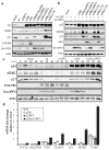
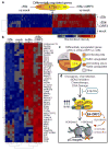
The transcription factor p53 (also known as TP53) guards against tumour and virus replication and is inactivated in almost all cancers. p53-activated transcription of target genes is thought to be synonymous with the stabilization of p53 in response to oncogenes and DNA damage. During adenovirus replication, the degradation of p53 by E1B-55k is considered essential for p53 inactivation, and is the basis for p53-selective viral cancer therapies. Here we reveal a dominant epigenetic mechanism that silences p53-activated transcription, irrespective of p53 phosphorylation and stabilization. We show that another adenoviral protein, E4-ORF3, inactivates p53 independently of E1B-55k by forming a nuclear structure that induces de novo H3K9me3 heterochromatin formation at p53 target promoters, preventing p53-DNA binding. This suppressive nuclear web is highly selective in silencing p53 promoters and operates in the backdrop of global transcriptional changes that drive oncogenic replication. These findings are important for understanding how high levels of wild-type p53 might also be inactivated in cancer as well as the mechanisms that induce aberrant epigenetic silencing of tumour-suppressor loci. Our study changes the longstanding definition of how p53 is inactivated in adenovirus infection and provides key insights that could enable the development of true p53-selective oncolytic viral therapies.
Figures


Comment in
-
Cancer: Viruses' backup plan.Nature. 2010 Aug 26;466(7310):1054-5. doi: 10.1038/4661054a. Nature. 2010. PMID: 20740004 No abstract available.
-
Cancer viruses: a two-pronged attack.Nat Rev Cancer. 2010 Oct;10(10):664. doi: 10.1038/nrc2935. Nat Rev Cancer. 2010. PMID: 21080563 No abstract available.
Similar articles
-
Late viral RNA export, rather than p53 inactivation, determines ONYX-015 tumor selectivity.Cancer Cell. 2004 Dec;6(6):611-23. doi: 10.1016/j.ccr.2004.11.012. Cancer Cell. 2004. PMID: 15607965
-
Impact of the adenoviral E4 Orf3 protein on the activity and posttranslational modification of p53.J Virol. 2015 Mar;89(6):3209-20. doi: 10.1128/JVI.03072-14. Epub 2015 Jan 7. J Virol. 2015. PMID: 25568206 Free PMC article.
-
Inhibition of p53 by adenovirus type 12 E1B-55K deregulates cell cycle control and sensitizes tumor cells to genotoxic agents.J Virol. 2011 Aug;85(16):7976-88. doi: 10.1128/JVI.00492-11. Epub 2011 Jun 15. J Virol. 2011. PMID: 21680522 Free PMC article.
-
p53 as a target for anti-cancer drug development.Crit Rev Oncol Hematol. 2006 Jun;58(3):190-207. doi: 10.1016/j.critrevonc.2005.10.005. Epub 2006 May 9. Crit Rev Oncol Hematol. 2006. PMID: 16690321 Review.
-
Interactions between adenovirus proteins and the p53 pathway: the development of ONYX-015.Semin Cancer Biol. 2000 Dec;10(6):453-9. doi: 10.1006/scbi.2000.0336. Semin Cancer Biol. 2000. PMID: 11170867 Review.
Cited by
-
p53-mediated heterochromatin reorganization regulates its cell fate decisions.Nat Struct Mol Biol. 2012 Apr 1;19(5):478-84, S1. doi: 10.1038/nsmb.2271. Nat Struct Mol Biol. 2012. PMID: 22466965 Free PMC article.
-
Human cytomegalovirus pUL29/28 and pUL38 repression of p53-regulated p21CIP1 and caspase 1 promoters during infection.J Virol. 2013 Mar;87(5):2463-74. doi: 10.1128/JVI.01926-12. Epub 2012 Dec 12. J Virol. 2013. PMID: 23236067 Free PMC article.
-
Timely synthesis of the adenovirus type 5 E1B 55-kilodalton protein is required for efficient genome replication in normal human cells.J Virol. 2012 Mar;86(6):3064-72. doi: 10.1128/JVI.06764-11. Epub 2012 Jan 25. J Virol. 2012. PMID: 22278242 Free PMC article.
-
Cancer viruses: a two-pronged attack.Nat Rev Cancer. 2010 Oct;10(10):664. doi: 10.1038/nrc2935. Nat Rev Cancer. 2010. PMID: 21080563 No abstract available.
-
Suppression of Type I Interferon Signaling by E1A via RuvBL1/Pontin.J Virol. 2017 Mar 29;91(8):e02484-16. doi: 10.1128/JVI.02484-16. Print 2017 Apr 15. J Virol. 2017. PMID: 28122980 Free PMC article.
References
-
- Levine AJ. The common mechanisms of transformation by the small DNA tumor viruses: The inactivation of tumor suppressor gene products: p53. Virology. 2008 - PubMed
-
- Lane DP, Crawford LV. T antigen is bound to a host protein in SV40-transformed cells. Nature. 1979;278:261–263. - PubMed
-
- Linzer DI, Levine AJ. Characterization of a 54K dalton cellular SV40 tumor antigen present in SV40-transformed cells and uninfected embryonal carcinoma cells. Cell. 1979;17:43–52. - PubMed
-
- Vogelstein B, Lane D, Levine AJ. Surfing the p53 network. Nature. 2000;408:307–310. - PubMed
-
- Vousden KH, Prives C. Blinded by the Light: The Growing Complexity of p53. Cell. 2009;137:413–431. - PubMed
Publication types
MeSH terms
Substances
Associated data
- Actions
Grants and funding
LinkOut - more resources
Full Text Sources
Other Literature Sources
Molecular Biology Databases
Research Materials
Miscellaneous

