

Comment
doi: 10.1038/nn0910-1045.
Zooming in on mouse vision
- PMID: 20740033
- PMCID: PMC3678908
- DOI: 10.1038/nn0910-1045
Item in Clipboard
Comment
Zooming in on mouse vision
Nat Neurosci.
2010 Sep.
Abstract
An examination of the micro-organization of visual cortex using two-photon calcium imaging provides a new level of insight into retinotopic maps, finding that retinotopy is scrambled on fine scales in mouse primary visual cortex.
Figures
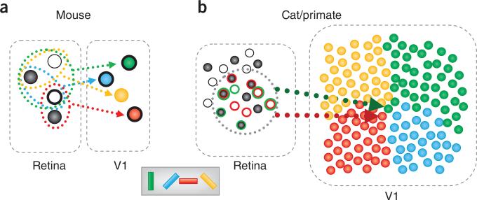
The mouse visual system is substantially different from those of cats and macaques. (a) In the mouse visual system, the retina contains about one RGC per input-layer neuron in V1. Some of the retinal ganglion cells respond to bright spots (ON center, white circles), whereas others respond to dark spots (OFF center, black circles). Neurons in mouse V1 are tuned for orientation (pictured as cell color) and receive input from a few RGCs. A shared subfield may be a result of common RGC input (heavy outline). The result of this arrangement is breakdown of retinotopy on a fine scale. Vertical (green) and horizontal (red) tuned cells share a subfield, despite being far from each other along cortex. (b) The situation in cats and macaques is quite different. In these systems, there are about 100 cortical recipient neurons for every RGC and these neurons are precisely arranged (for example, by their orientation preference, as illustrated for an orientation pinwheel on the right). Given that many RGCs contribute to a single receptive field in V1, whereas each RGC diverges to contribute to the receptive fields of multiple V1 neurons, many orientations may be adequately represented without the apparent need to sacrifice retinotopy. Compare the inputs into a cell tuned for horizontal (red) versus vertical (green). The net receptive field position of these cells would be quite similar (dotted circle). Other retinotopic positions may be smoothly mapped by sliding the dotted outline. Outline colors of RGCs indicate which RGCs connect to which neuron (those outlined in green connect to the green neuron and similarly for red).
Comment on
-
Parallel processing of visual space by neighboring neurons in mouse visual cortex.Nat Neurosci. 2010 Sep;13(9):1144-9. doi: 10.1038/nn.2620. Epub 2010 Aug 15. Nat Neurosci. 2010. PMID: 20711183 Free PMC article.
References
Publication types
MeSH terms
Grants and funding
LinkOut - more resources
Full Text Sources
