

doi: 10.3390/v2051146.
The Role of Lipids in Retrovirus Replication
Affiliations
- PMID: 20740061
- PMCID: PMC2927015
- DOI: 10.3390/v2051146
Item in Clipboard
The Role of Lipids in Retrovirus Replication
Viruses.
.
Abstract
Retroviruses undergo several critical steps to complete a replication cycle. These include the complex processes of virus entry, assembly, and budding that often take place at the plasma membrane of the host cell. Both virus entry and release involve membrane fusion/fission reactions between the viral envelopes and host cell membranes. Accumulating evidence indicates important roles for lipids and lipid microdomains in virus entry and egress. In this review, we outline the current understanding of the role of lipids and membrane microdomains in retroviral replication.
Figures
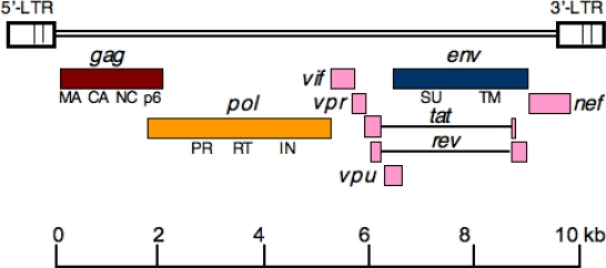
Genomic organization of HIV-1. The rectangles indicate the open reading frames for the gag (brown), pol (yellow), and env (blue) genes. Complex retroviruses like HIV-1 encode additional regulatory and accessory genes (shown in pink). Tat and Rev introns are depicted as horizontal lines. The major Gag domains matrix (MA), capsid (CA), nucleocapsid (NC), and p6 and the Env surface (SU) and transmembrane (TM) glycoproteins (gp120 and gp41, respectively, in the case of HIV-1) are indicated. The 5′ and 3′ long terminal repeats (LTRs) are also shown.
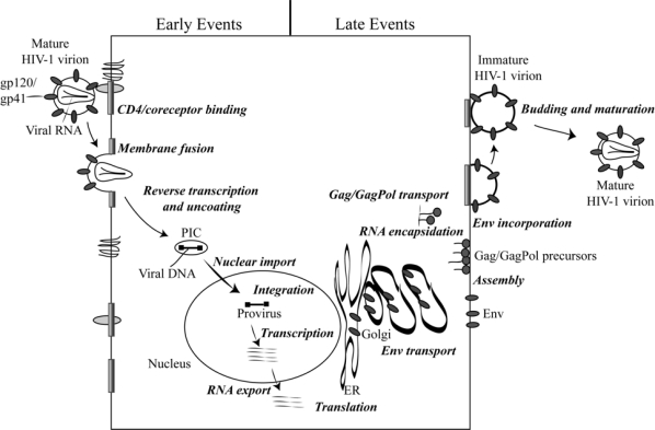
Schematic representation of the HIV-1 replication cycle. The steps that comprise the replication cycle are described in more detail in the text. Reprinted with permission from Freed [221]. Copyright 2004 Elsevier Inc.
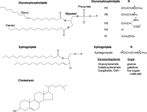
Structures of major classes of lipids in eukaryotic cells. Glycerophospholipids contain diacylglycerol, a phosphate group, and a simple organic head group. The acyl chains contain 16–18 carbon atoms and one of the acyl chains is generally unsaturated with a cis double bond. The head groups vary among the phospholipids; e.g., choline in phosphatidylcholine (PC), ethanolamine in phosphatidylethanolamine (PE), serine in phosphatidylserine (PS), and inositol in phosphatidylinositol (PI). Sphingolipids are based on ceramide, which has long saturated acyl chains of 16–26 carbon atoms, and either phosphorylcholine (sphingomyelin) or sugar (glycosphingolipids) headgroups. Depending on the type of sugar, glycosphingolipids are further sub-classified into several types as listed in the figure. Cholesterol is a major sterol in mammalian cells.
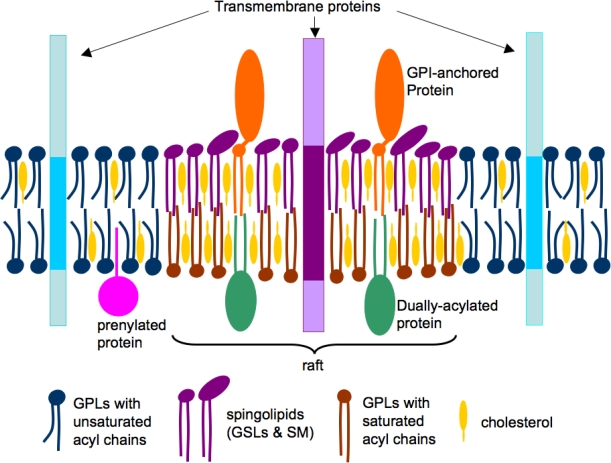
A simplified model of lipid rafts in cell membranes. The phospholipids (blue and brown) and cholesterol (yellow) are distributed in both the leaflets, whereas sphingolipids (violet) are enriched in the outer leaflet of the bilayer. The acyl chains of raft lipids are generally long and saturated (violet and brown), whereas those in non-raft domains are shorter and contain singly or multiply unsaturated acyl chains (blue). Raft domains contain concentrations of dually-acylated (green) and GPI-anchored (brown) proteins, whereas transmembrane (blue) and prenylated (green) proteins are usually non-raft associated. Reprinted with permission from Waheed and Freed [2]. Copyright 2009. Elsevier Inc.
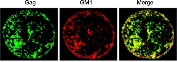
Copatching of HIV-1 Gag proteins with the raft marker GM1. Before fixation, Jurkat cells expressing HIV-1 Gag were treated with cholera toxin B subunit to stain GM1. Cells were then fixed, permeabilized, and Gag proteins were detected with an anti-p17 MA antibody. Gag and GM1 show colocalization, as shown in yellow in the merged image. Reprinted with permission from Ono and Freed [1]. Copyright 2005, Elsevier Inc.
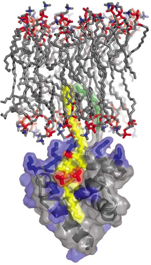
Complex between HIV-1 MA and PI(4,5)P2. The highly basic surface of MA (blue) exhibits electrostatic interactions with PI(4,5)P2 (yellow and red phosphates). The 2′-unsaturated acyl chain of PI(4,5)P2 (yellow) binds to the hydrophobic cleft in MA and the myristyl group (green) of MA inserts into the lipid bilayer. Unpublished image provided by Dr. M. Summers, based on the data of Saad et al. [124].
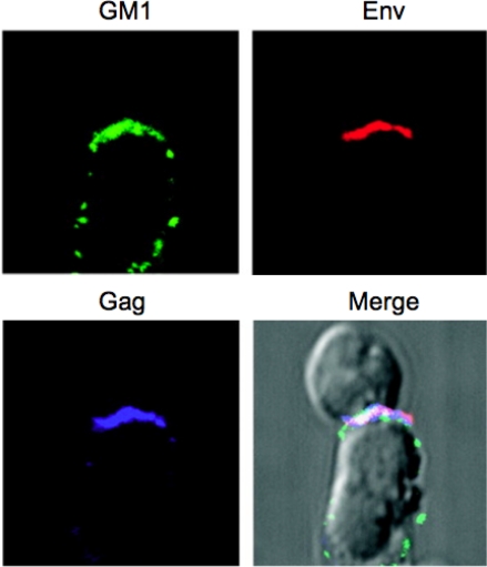
Virological synapse showing HIV-1 Gag and Env concentrating in a raft-rich domain. Primary CD4+ T-cells (top) were incubated with HIV-1-infected Jurkat cells (bottom) for 1 h at 37 °C. During the incubation, cells were stained for Env (red), fixed and stained with cholera toxin B subunit (green), specific for the raft marker GM1. Cells were permeabilized and Gag proteins were detected with anti-p17 antibody (blue). Images were superimposed; white indicates the colocalization of three colors. Reprinted with permission from Jolly and Sattentau [191]. Copyright 2005, The American Society of Microbiology.
Similar articles
-
Lipids and membrane microdomains in HIV-1 replication.Virus Res. 2009 Aug;143(2):162-76. doi: 10.1016/j.virusres.2009.04.007. Epub 2009 Apr 19. Virus Res. 2009. PMID: 19383519 Free PMC article. Review.
-
Virus infection and lipid rafts.Biol Pharm Bull. 2006 Aug;29(8):1538-41. doi: 10.1248/bpb.29.1538. Biol Pharm Bull. 2006. PMID: 16880600 Review.
-
Role of lipids in virus replication.Cold Spring Harb Perspect Biol. 2011 Oct 1;3(10):a004820. doi: 10.1101/cshperspect.a004820. Cold Spring Harb Perspect Biol. 2011. PMID: 21628428 Free PMC article. Review.
-
Host Cell Plasma Membrane Phosphatidylserine Regulates the Assembly and Budding of Ebola Virus.J Virol. 2015 Sep;89(18):9440-53. doi: 10.1128/JVI.01087-15. Epub 2015 Jul 1. J Virol. 2015. PMID: 26136573 Free PMC article.
-
Assembly and budding of influenza virus.Virus Res. 2004 Dec;106(2):147-65. doi: 10.1016/j.virusres.2004.08.012. Virus Res. 2004. PMID: 15567494 Free PMC article. Review.
Cited by
-
How HIV finds the door.Proc Natl Acad Sci U S A. 2012 Nov 13;109(46):18631-2. doi: 10.1073/pnas.1215940109. Epub 2012 Nov 1. Proc Natl Acad Sci U S A. 2012. PMID: 23118338 Free PMC article. No abstract available.
-
The lipid membrane of HIV-1 stabilizes the viral envelope glycoproteins and modulates their sensitivity to antibody neutralization.J Biol Chem. 2020 Jan 10;295(2):348-362. doi: 10.1074/jbc.RA119.009481. Epub 2019 Nov 22. J Biol Chem. 2020. PMID: 31757809 Free PMC article.
-
Global rescue of defects in HIV-1 envelope glycoprotein incorporation: implications for matrix structure.PLoS Pathog. 2013;9(11):e1003739. doi: 10.1371/journal.ppat.1003739. Epub 2013 Nov 14. PLoS Pathog. 2013. PMID: 24244165 Free PMC article.
-
New insights into HIV assembly and trafficking.Physiology (Bethesda). 2011 Aug;26(4):236-51. doi: 10.1152/physiol.00051.2010. Physiology (Bethesda). 2011. PMID: 21841072 Free PMC article. Review.
-
Trio engagement via plasma membrane phospholipids and the myristoyl moiety governs HIV-1 matrix binding to bilayers.Proc Natl Acad Sci U S A. 2013 Feb 26;110(9):3525-30. doi: 10.1073/pnas.1216655110. Epub 2013 Feb 11. Proc Natl Acad Sci U S A. 2013. PMID: 23401539 Free PMC article.
References
-
- Ono A, Freed EO. Role of lipid rafts in virus replication. Adv Virus Res. 2005;64:311–358. - PubMed
-
- Freed E, Martin M. HIVs and their replication. In: Knipe D, Howley P, editors. Fields Virology. Lippincott; Williams, and Wilkins, USA: 2007. pp. 2107–2185.
-
- Munro S. Lipid rafts: elusive or illusive. Cell. 2003;115:377–388. - PubMed
-
- Simons K, Vaz WL. Model systems, lipid rafts, and cell membranes. Annu Rev Biophys Biomol Struct. 2004;33:269–295. - PubMed
Grants and funding
LinkOut - more resources
Full Text Sources
Other Literature Sources