

Functional connectivity of cortical motor areas in the resting state in Parkinson's disease
- PMID: 20740649
- PMCID: PMC6870250
- DOI: 10.1002/hbm.21118
Functional connectivity of cortical motor areas in the resting state in Parkinson's disease
Abstract
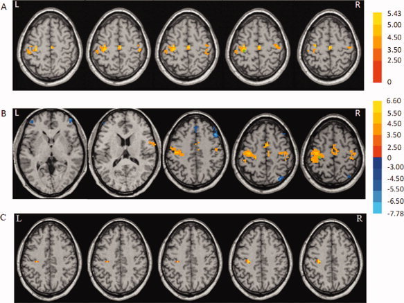
Parkinson's disease (PD) patients have difficulty in initiating movements. Previous studies have suggested that the abnormal brain activity may happen not only during performance of self-initiated movements but also in the before movement (baseline or resting) state. In the current study, we investigated the functional connectivity of brain networks in the resting state in PD. We chose the rostral supplementary motor area (pre-SMA) and bilateral primary motor cortex (M1) as "seed" regions, because the pre-SMA is important in motor preparation, whereas the M1 is critical in motor execution. FMRIs were acquired in 18 patients and 18 matched controls. We found that in the resting state, the pattern of connectivity with both the pre-SMA or the M1 was changed in PD. Connectivity with the pre-SMA in patients with PD compared to normal subjects was increased connectivity to the right M1 and decreased to the left putamen, right insula, right premotor cortex, and left inferior parietal lobule. We only found stronger connectivity in the M1 with its own local region in patients with PD compared to controls. Our findings demonstrate that the interactions of brain networks are abnormal in PD in the resting state. There are more connectivity changes of networks related to motor preparation and initiation than to networks of motor execution in PD. We postulate that these disrupted connections indicate a lack of readiness for movement and may be partly responsible for difficulty in initiating movements in PD.
Copyright © 2010 Wiley-Liss, Inc.
Figures


References
-
- Alexander GE,Crutcher MD,DeLong MR ( 1990): Basal ganglia‐thalamocortical circuits: Parallel substrates for motor, oculomotor, “prefrontal” and “limbic” functions. Progr Brain Res 85: 119–146. - PubMed
-
- Augustine JR ( 1996): Circuitry and functional aspects of the insular lobe in primates including humans. Brain Res Rev 22: 229–244. - PubMed
-
- Bares M,Kanovsky P,Klajblova H,Rektor I ( 2003): Intracortical inhibition and facilitation are impaired in patients with early Parkinson's disease: A paired TMS study. Eur J Neurol 10: 385–389. - PubMed
-
- Benamer HT,Oertel WH,Patterson J,Hadley DM,Pogarell O,Hoffken H,Gerstner A,Grosset DG ( 2003): Prospective study of presynaptic dopaminergic imaging in patients with mild parkinsonism and tremor disorders: Part 1. Baseline and 3‐month observations. Mov Disord 18: 977–984. - PubMed
-
- Birn RM,Diamond JB,Smith MA,Bandettini PA ( 2006): Separating respiratory‐variation‐related fluctuations from neuronal‐activity‐related fluctuations in fMRI. Neuroimage 31: 1536–1548. - PubMed
Publication types
MeSH terms
Substances
LinkOut - more resources
Full Text Sources
Other Literature Sources
Medical
