

Determination of spin compartment in arterial spin labeling MRI
- PMID: 20740655
- PMCID: PMC2994958
- DOI: 10.1002/mrm.22601
Determination of spin compartment in arterial spin labeling MRI
Abstract
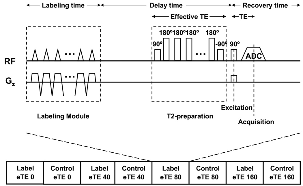
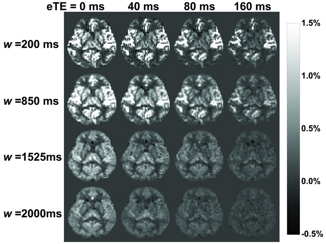
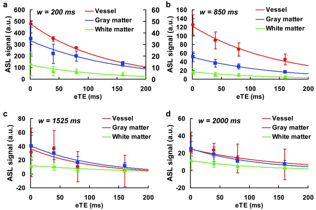
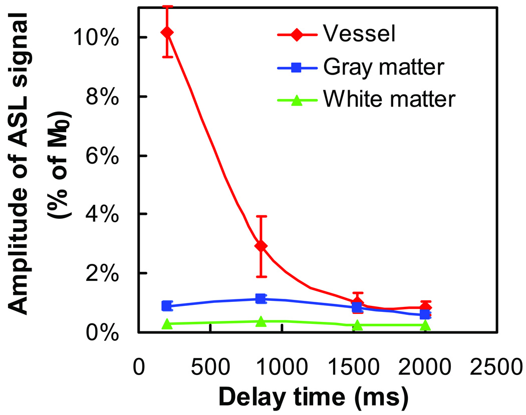
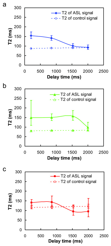
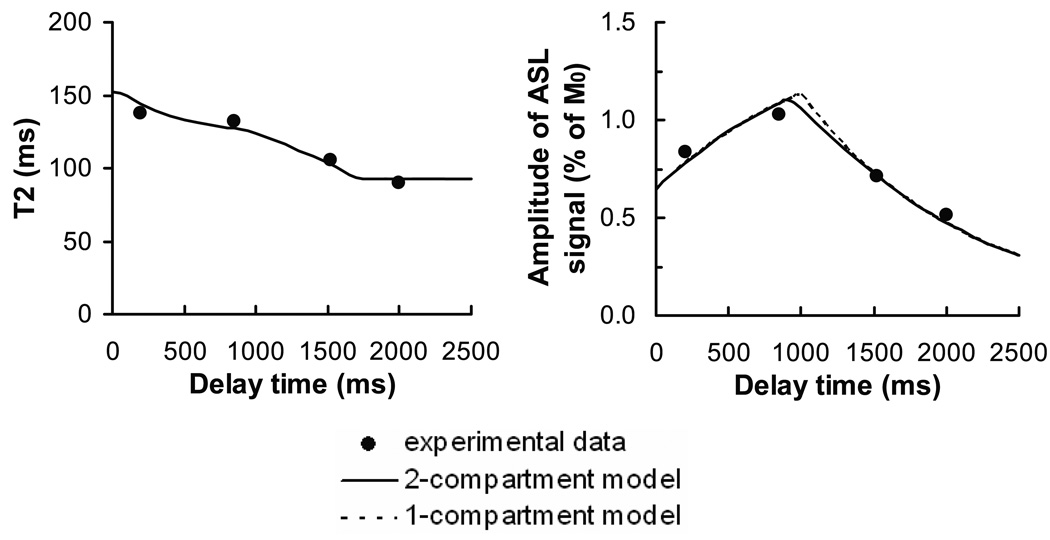
A major difference between arterial-spin-labeling MRI and gold-standard radiotracer blood flow methods is that the compartment localization of the labeled spins in the arterial-spin-labeling image is often ambiguous, which may affect the quantification of cerebral blood flow. In this study, we aim to probe whether the spins are located in the vascular system or tissue by using T2 of the arterial-spin-labeling signal as a marker. We combined two recently developed techniques, pseudo-continuous arterial spin labeling and T2-Relaxation-Under-Spin-Tagging, to determine the T2 of the labeled spins at multiple postlabeling delay times. Our data suggest that the labeled spins first showed the T2 of arterial blood followed by gradually approaching and stabilizing at the tissue T2. The T2 values did not decrease further toward the venous T2. By fitting the experimental data to a two-compartment model, we estimated gray matter cerebral blood flow, arterial transit time, and tissue transit time to be 74.0 ± 10.7 mL/100g/min (mean ± SD, N = 10), 938 ± 156 msec, and 1901 ± 181 msec, respectively. The arterial blood volume was calculated to be 1.18 ± 0.21 mL/100 g. A postlabeling delay time of 2 s is sufficient to allow the spins to completely enter the tissue space for gray matter but not for white matter.
© 2010 Wiley-Liss, Inc.
Figures

References
-
- Mintun MA, Raichle ME, Martin WR, Herscovitch P. Brain oxygen utilization measured with O-15 radiotracers and positron emission tomography. J Nucl Med. 1984;25(2):177–187. - PubMed
-
- Alsop DC, Detre JA. Reduced transit-time sensitivity in noninvasive magnetic resonance imaging of human cerebral blood flow. J Cereb Blood Flow Metab. 1996;16(6):1236–1249. - PubMed
-
- Buxton RB, Frank LR, Wong EC, Siewert B, Warach S, Edelman RR. A general kinetic model for quantitative perfusion imaging with arterial spin labeling. Magn Reson Med. 1998;40(3):383–396. - PubMed
-
- Gonzalez-At JB, Alsop DC, Detre JA. Cerebral perfusion and arterial transit time changes during task activation determined with continuous arterial spin labeling. Magn Reson Med. 2000;43(5):739–746. - PubMed
Publication types
MeSH terms
Substances
Grants and funding
LinkOut - more resources
Full Text Sources
