

Strain and torsion quantification in mouse hearts under dobutamine stimulation using 2D multiphase MR DENSE
- PMID: 20740659
- PMCID: PMC2965822
- DOI: 10.1002/mrm.22530
Strain and torsion quantification in mouse hearts under dobutamine stimulation using 2D multiphase MR DENSE
Abstract
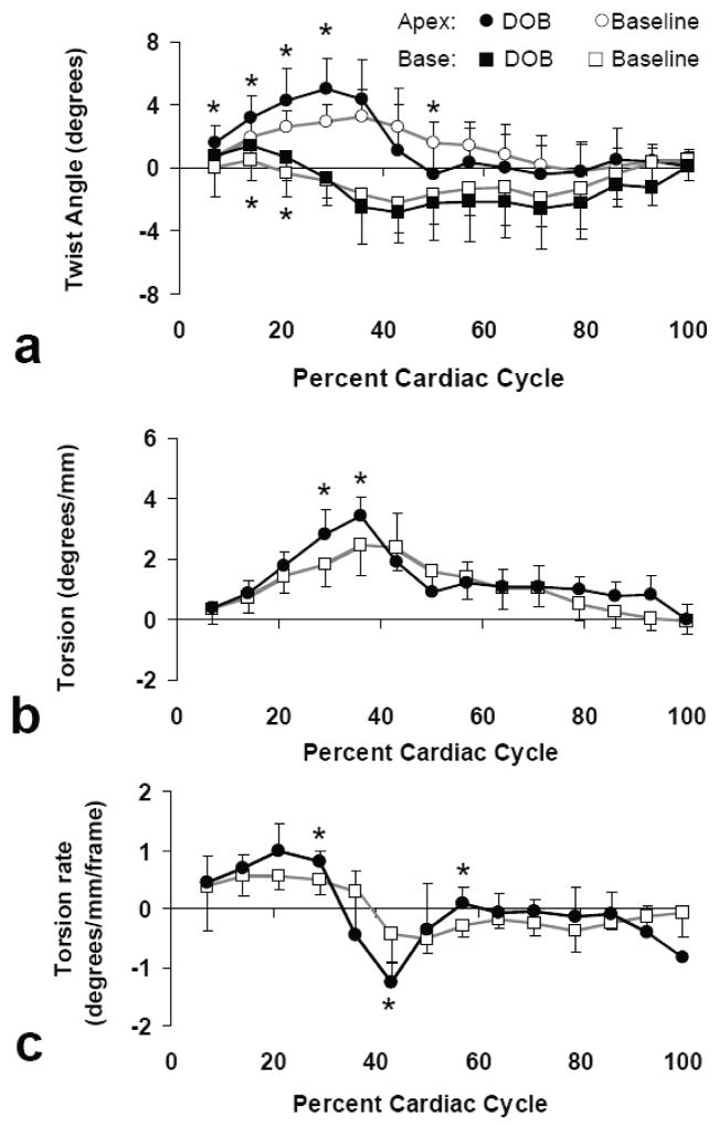
In this study, a 2D multiphase magnetic resonance displacement encoding with stimulated echoes (DENSE) imaging and analysis method was developed for direct quantification of Lagrangian strain in the mouse heart. Using the proposed method, <10 ms temporal resolution and 0.56 mm in-plane resolution were achieved. A validation study that compared strain calculation by displacement encoding with stimulated echoes and by magnetic resonance tagging showed high correlation between the two methods (R(2) > 0.80). Regional ventricular wall strain and twist were characterized in mouse hearts at baseline and under dobutamine stimulation. Dobutamine stimulation induced significant increase in radial and circumferential strains and torsion at peak systole. A rapid untwisting was also observed during early diastole. This work demonstrates the capability of characterizing cardiac functional response to dobutamine stimulation in the mouse heart using 2D multiphase magnetic resonance displacement encoding with stimulated echoes.
Copyright © 2010 Wiley-Liss, Inc.
Figures


Similar articles
-
Myocardial 3D strain calculation by combining cine displacement encoding with stimulated echoes (DENSE) and cine strain encoding (SENC) imaging.Magn Reson Med. 2009 Jul;62(1):77-84. doi: 10.1002/mrm.21984. Magn Reson Med. 2009. PMID: 19322795 Free PMC article.
-
Imaging three-dimensional myocardial mechanics using navigator-gated volumetric spiral cine DENSE MRI.Magn Reson Med. 2010 Oct;64(4):1089-97. doi: 10.1002/mrm.22503. Magn Reson Med. 2010. PMID: 20574967 Free PMC article.
-
Off-resonance insensitive complementary SPAtial Modulation of Magnetization (ORI-CSPAMM) for quantification of left ventricular twist.J Magn Reson Imaging. 2014 Feb;39(2):339-45. doi: 10.1002/jmri.24154. Epub 2013 Apr 26. J Magn Reson Imaging. 2014. PMID: 23625854 Free PMC article.
-
Optimal configuration of respiratory navigator gating for the quantification of left ventricular strain using spiral cine displacement encoding with stimulated echoes (DENSE) MRI.J Magn Reson Imaging. 2017 Mar;45(3):786-794. doi: 10.1002/jmri.25389. Epub 2016 Jul 26. J Magn Reson Imaging. 2017. PMID: 27458823 Free PMC article.
-
Ventricular mechanics: techniques and applications.Magn Reson Imaging Clin N Am. 2015 Feb;23(1):7-13. doi: 10.1016/j.mric.2014.08.005. Magn Reson Imaging Clin N Am. 2015. PMID: 25476669 Review.
Cited by
-
Myocardial tagging by cardiovascular magnetic resonance: evolution of techniques--pulse sequences, analysis algorithms, and applications.J Cardiovasc Magn Reson. 2011 Jul 28;13(1):36. doi: 10.1186/1532-429X-13-36. J Cardiovasc Magn Reson. 2011. PMID: 21798021 Free PMC article. Review.
-
Inducible re-expression of HEXIM1 causes physiological cardiac hypertrophy in the adult mouse.Cardiovasc Res. 2013 Jul 1;99(1):74-82. doi: 10.1093/cvr/cvt086. Epub 2013 Apr 11. Cardiovasc Res. 2013. PMID: 23585471 Free PMC article.
-
The Structural Basis of Functional Improvement in Response to Human Umbilical Cord Blood Stem Cell Transplantation in Hearts With Postinfarct LV Remodeling.Cell Transplant. 2015;24(6):971-83. doi: 10.3727/096368913X675746. Epub 2013 Dec 10. Cell Transplant. 2015. PMID: 24332083 Free PMC article.
-
Inter-study reproducibility of left ventricular torsion and torsion rate quantification using MR myocardial feature tracking.J Magn Reson Imaging. 2016 Jan;43(1):128-37. doi: 10.1002/jmri.24979. Epub 2015 Jun 26. J Magn Reson Imaging. 2016. PMID: 26114731 Free PMC article.
-
Quantification of myocardial strain in patients with isolated left ventricular non-compaction and healthy subjects using deformable registration algorithm: comparison with feature tracking.BMC Cardiovasc Disord. 2020 Sep 3;20(1):400. doi: 10.1186/s12872-020-01668-x. BMC Cardiovasc Disord. 2020. PMID: 32883201 Free PMC article.
References
-
- James JF, Hewett TE, Robbins J. Cardiac physiology in transgenic mice. Circ Res. 1998;82:407–415. - PubMed
-
- Zerhouni EA, Parish DM, Rogers WJ, Yang A, Shapiro EP. Human heart: tagging with MR imaging--a method for noninvasive assessment of myocardial motion. Radiology. 1988;169:59–63. - PubMed
-
- Axel L, Dougherty L. MR imaging of motion with spatial modulation of magnetization. Radiology. 1989;171:841–845. - PubMed
-
- Axel L, Dougherty L. Heart wall motion: improved method of spatial modulation of magnetization for MR imaging. Radiology. 1989;172:349–350. - PubMed
-
- Zhou R, Pickup S, Glickson JD, Scott CH, Ferrari VA. Assessment of global and regional myocardial function in the mouse using cine and tagged MRI. Magn Reson Med. 2003;49:760–764. - PubMed
Publication types
MeSH terms
Substances
Grants and funding
LinkOut - more resources
Full Text Sources
