. 2011 Feb;29(2):289-96.
doi: 10.1002/jor.21220. Epub 2010 Aug 25.

Scleraxis expression is coordinately regulated in a murine model of patellar tendon injury

Affiliations

Scleraxis expression is coordinately regulated in a murine model of patellar tendon injury

Alexander Scott et al. J Orthop Res. 2011 Feb.

Abstract

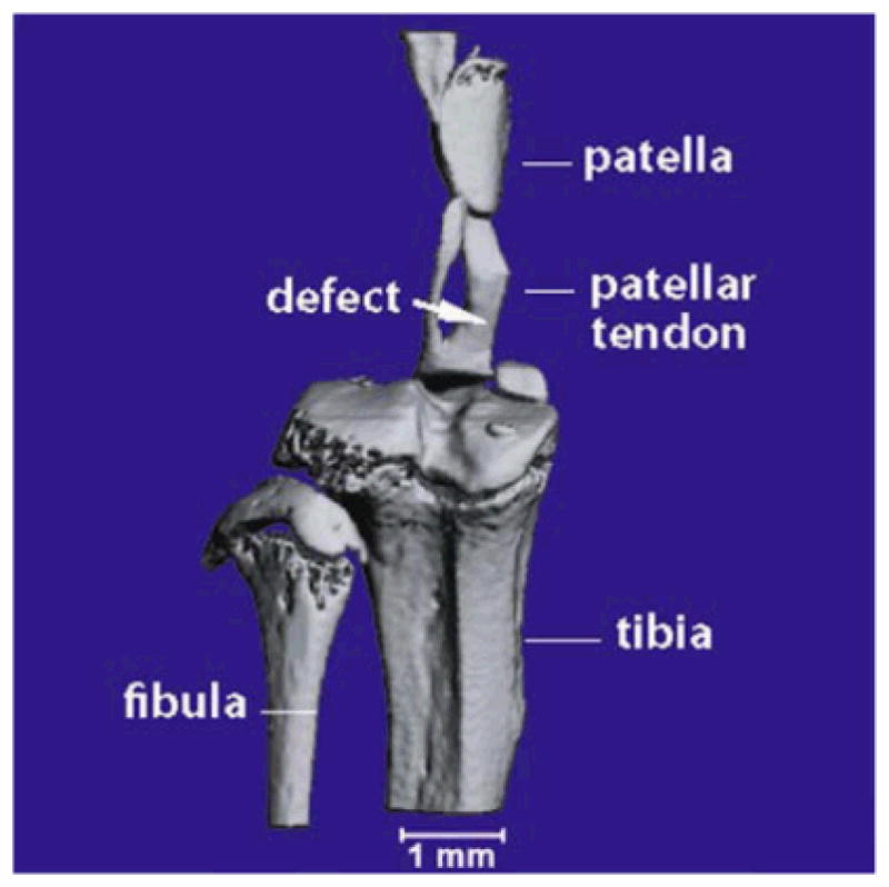
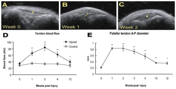
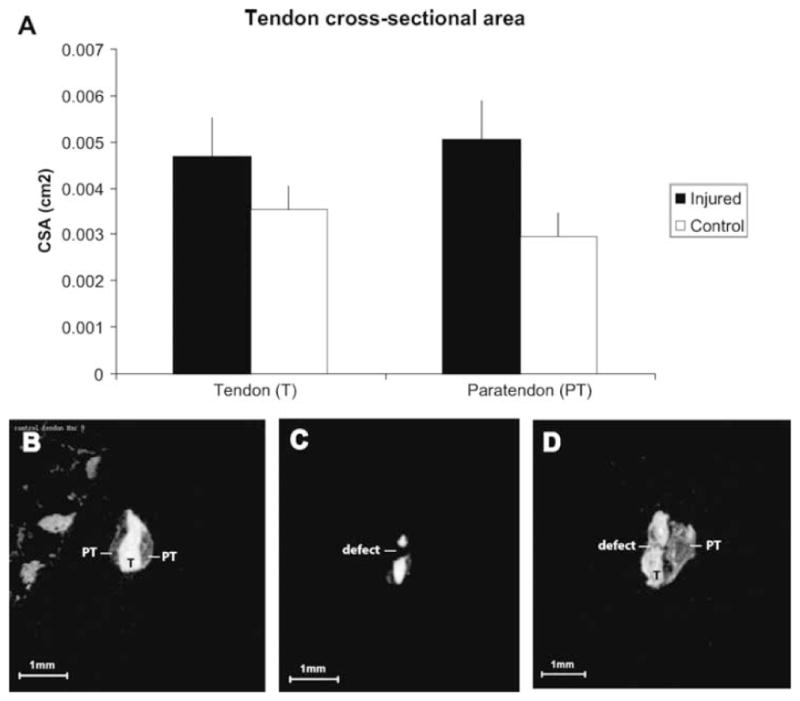
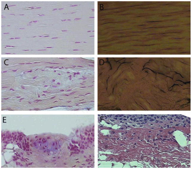
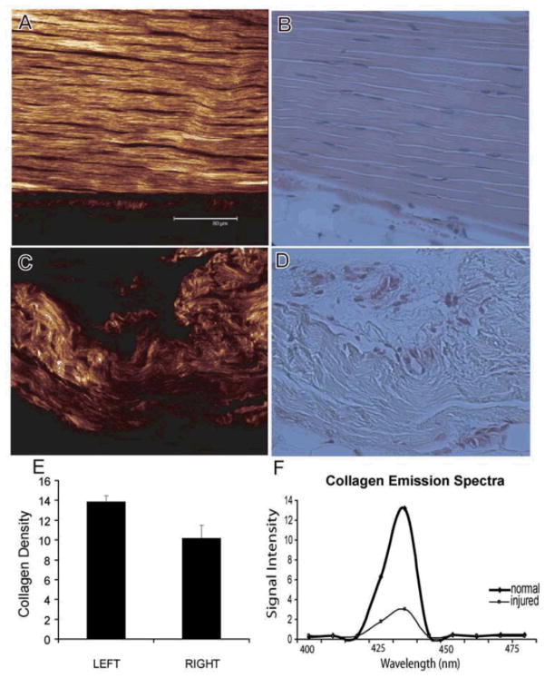
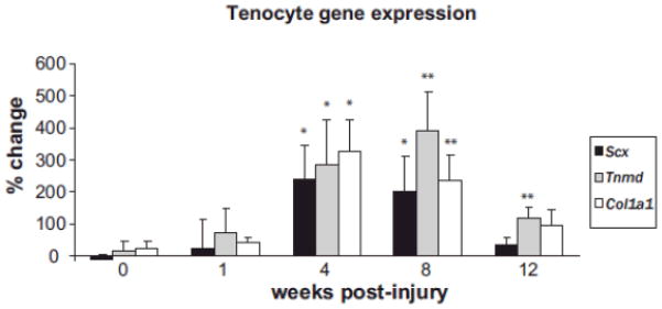
This study investigated the expression of Scleraxis in a murine model of patellar tendon injury in which the central third of the patellar tendon was unilaterally injured. The presence of tendon pathology was assessed using dual photon microscopy, conventional histology and microCT. Tendon pathology was also quantified noninvasively over a 12-week period using high-frequency ultrasound and laser Doppler flowmetry. Gene expression (Scx, Tnmd, and Col1a1) was determined at defined end-points (1, 4, 8, and 12 weeks) using qPCR on RNA from individual patellar tendons on injured and uninjured sides. There was significant development of tendon pathology as gauged by ultrasound and laser Doppler over 12 weeks. Injured tendons demonstrated significant histological and microCT evidence of pathological change, and disorganized collagen with reduced density. The expression of Scx and Col1a1 was unchanged at 1 week, significantly upregulated at 4 and 8 weeks, and had returned to baseline by 12 weeks. Tnmd expression was unchanged at 1 week, and significantly increased at 4, 8, and 12 weeks. Patellar tendon injury was associated with marked increases in the expression of Scx, Tnmd, and Col1a1. Our data suggest new roles for Scleraxis in coordinating the response to injury in the pathogenesis of tendon disorders.


Figures

Figure 1
Figure 1
MicroCT of right mouse patellar tendon. A representative scan illustrating the nature and location of the central defect immediately following injury.
Figure 2
Figure 2
Noninvasive measurement of patellar tendon pathology. Top panels (A–C) demonstrate evolving ultrasound appearance of a mouse patellar tendon before (week 0) and after (weeks 1, 2) injury. (A) Tendon denoted by asterisk. (B) Hypoechoic area corresponding to sight of defect marked with arrowhead. (C) Tendon marked by asterisk—note substantial tendon thickening. (D,E) Tendon thickness and blood flow gradually decreased over time but remained significantly increased at 12 weeks (p<0.001 for thickness, p=0.0312 for blood flow).
Figure 3
Figure 3
MicroCT analysis of tendon and paratendon. (A) At 4 weeks, both tendon and paratendon were significantly greater in crosssectional area (CSA). *p<0.05. Lower panels: Representative microCT images of tendon that is uninjured (B), immediately post-injury (C), and 3 days following injury (D). In D, substantial thickening can be appreciated both in the tendon (T) and paratendon (PT). Note the in homogenous texture of tendon and paratendon, as well as the irregularity of tendon and paratendon boundaries.
Figure 4
Figure 4
Histology of injured and uninjured tendon. Uninjured tendon demonstrated regular cellularity (A, fast nuclear red [FNR]) and ordered collagen (B, picrosirius red [PSR]). Sixteen weeks following injury, areas of cellular (C, FNR) and collagen (D, PSR) disarray were prevalent. Other abnormalities present in injured, but not in control, tendon included fibrocartilage metaplasia (E, FNR and Alcian Blue) and expansion of paratendon (F, H&E). In E, note the area of glycosaminoglycan accumulation (blue staining) surrounding chondrocyte-like cells. All images at same scale, original magnification, ×400.
Figure 5
Figure 5
Representative second harmonic generation (SHG: A,C) and corresponding histological images (H&E: B,D) of normal and injured tendon tissues. The scale bar in A is 80.0 _m, and all images are at the same scale. (E)We observe that the collagen density in the active repair area is significantly reduced compared to uninjured tendons (p=0.004). (F) A representative histogram of the detected SHG signal demonstrates the distribution of the collagen molecular density obtained from normal tendon as well as the one undergoing active repair. The detected SHG signal is clearly manifested by a narrow peak and is spectrally clean, arising only at the expected wavelength of 440 nm. Considerable differences in the collagen matrix are observed between the normal and injured tendon tissues.
Figure 6
Figure 6
Gene expression in injured patellar tendon. Values are normalized to 18S rRNA, and compared to control patellar tendon from the same animal and expressed as % change. Scx, Scleraxis; Tnmd, tenomodulin; Col1a1, collagen type 1a1. *p<0.0.05, **p<0.01. n=8 tendons per data point.

Similar articles

Cited by

References

    1. Hazleman B, Riley G, Speed CA. Soft tissue rheumatology. Oxford: Oxford University Press; 2004.
    1. Tallon C, Coleman BD, Khan KM, et al. Outcome of surgery for chronic achilles tendinopathy. A critical review. Am J Sports Med. 2001;29:315–320. - PubMed
    1. Cook JL, Khan KM, Harcourt PR, et al. A cross sectional study of 100 athletes with jumper’s knee managed conservatively and surgically. The Victorian Institute of Sport Tendon Study Group. Br J Sports Med. 1997;31:332– 336. - PMC - PubMed
    1. Maffulli N, Wong J, Almekinders LC. Types and epidemiology of tendinopathy. Clin Sports Med. 2003;22:675–692. - PubMed
    1. Jarvinen M, Jozsa L, Kannus P, et al. Histopathological findings in chronic tendon disorders. Scand JMed Sci Sports. 1997;7:86–95. - PubMed

Publication types

Grants and funding