

Memory T cells in Rhesus macaques
- PMID: 20795545
- PMCID: PMC7316376
- DOI: 10.1007/978-1-4419-6451-9_10
Memory T cells in Rhesus macaques
Abstract
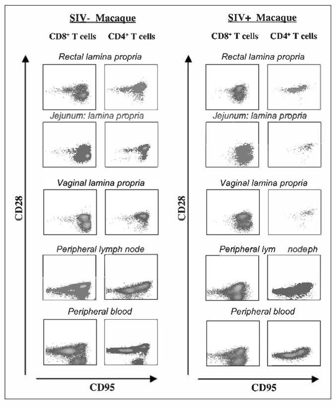
The Rhesus macaque (Macaca mulatta) is one of the best studied species of Old World monkeys. DNA sequencing of the entire Rhesus macaque genome, completed in 2007, has demonstrated that humans and macaques share about 93% of their nucleotide sequence. Rhesus macaques have been widely used for medical research including drug testing, neurology, behavioral and cognitive science, reproduction, xenotransplantation and genetics. Because of the Rhesus macaque's sensitivity to bacteria, parasites and viruses that cause similar disease in humans, these animals represent an excellent model to study infectious diseases. The recent pandemic of HIV and the discovery of SIV, a lentivirus genetically related to HIV Type 1 that causes AIDS in Rhesus macaques, have prompted the development of reagents that can be used to study innate and adaptive immune responses in macaques at the single cell level. This review will focus on the distribution of memory cells in the different immunologic compartments of Rhesus macaques. In addition, the strategies available to manipulate memory cells in Rhesus macaques to understand their trafficking and function will be discussed. Emphasis is placed on studies of memory cells in macaques infected with SIV because many studies are available. Lastly, we highlight the usefulness of the Rhesus macaque model in studies related to the aging of the immune system.
Figures
Similar articles
-
Aged Chinese rhesus macaques suffer severe phenotypic T- and B-cell aging accompanied with sex differences.Exp Gerontol. 2014 Jul;55:113-9. doi: 10.1016/j.exger.2014.04.004. Epub 2014 Apr 16. Exp Gerontol. 2014. PMID: 24746513
-
Initiation of Antiretroviral Therapy Restores CD4+ T Memory Stem Cell Homeostasis in Simian Immunodeficiency Virus-Infected Macaques.J Virol. 2016 Jul 11;90(15):6699-6708. doi: 10.1128/JVI.00492-16. Print 2016 Aug 1. J Virol. 2016. PMID: 27170752 Free PMC article.
-
Elite Control, Gut CD4 T Cell Sparing, and Enhanced Mucosal T Cell Responses in Macaca nemestrina Infected by a Simian Immunodeficiency Virus Lacking a gp41 Trafficking Motif.J Virol. 2015 Oct;89(20):10156-75. doi: 10.1128/JVI.01134-15. Epub 2015 Jul 29. J Virol. 2015. PMID: 26223646 Free PMC article.
-
Immunologic and pathologic manifestations of the infection of rhesus monkeys with simian immunodeficiency virus of macaques.J Acquir Immune Defic Syndr (1988). 1990;3(11):1023-40. J Acquir Immune Defic Syndr (1988). 1990. PMID: 2213505 Review.
-
Immune senescence in aged nonhuman primates.Exp Gerontol. 2010 Sep;45(9):655-61. doi: 10.1016/j.exger.2010.06.001. Epub 2010 Jun 15. Exp Gerontol. 2010. PMID: 20558288 Free PMC article. Review.
Cited by
-
Safety and Immunogenicity Study of a Bivalent Vaccine for Combined Prophylaxis of COVID-19 and Influenza in Non-Human Primates.Vaccines (Basel). 2024 Sep 26;12(10):1099. doi: 10.3390/vaccines12101099. Vaccines (Basel). 2024. PMID: 39460266 Free PMC article.
-
Nonhuman primate models of type 1 diabetes mellitus for islet transplantation.J Diabetes Res. 2014;2014:785948. doi: 10.1155/2014/785948. Epub 2014 Oct 20. J Diabetes Res. 2014. PMID: 25389531 Free PMC article. Review.
-
Decidual leukocytes respond to African lineage Zika virus infection with mild anti-inflammatory changes during acute infection in rhesus macaques.Front Immunol. 2024 Mar 7;15:1363169. doi: 10.3389/fimmu.2024.1363169. eCollection 2024. Front Immunol. 2024. PMID: 38515747 Free PMC article.
-
Long-Term Recovery of the Adaptive Immune System in Rhesus Macaques After Total Body Irradiation.Adv Radiat Oncol. 2021 Apr 19;6(5):100677. doi: 10.1016/j.adro.2021.100677. eCollection 2021 Sep-Oct. Adv Radiat Oncol. 2021. PMID: 34646962 Free PMC article.
-
Belatacept-Resistant Rejection Is Associated With CD28+ Memory CD8 T Cells.Am J Transplant. 2017 Sep;17(9):2285-2299. doi: 10.1111/ajt.14349. Epub 2017 Jul 11. Am J Transplant. 2017. PMID: 28502128 Free PMC article.
References
-
- Picker LJ, Siegelman MH. Lymphoid organs and tissues In: Paul WE, ed. Fundamental Immunology, 4th ed. Philadelphia: Lippincott-Raven, 1999:14, 479.
-
- Picker LJ, Terstappen LW, Rott LS et al. Differential expression of homing-associated adhesion molecules by T-cell subsets in man. J Immunol 1990; 145:3247–3255. - PubMed
-
- Mestas J, Hughes CCW. Of mice and not men: differences between mouse and human immunology. J Immunol 2004; 172:2731–2738. - PubMed
-
- ACLAM [American College of Laboratory Animal Medicine]. Public statement: medical records for animals used in research, teaching and testing. ILAR journal 2007; 48(1). Public statement. - PubMed
-
- Conlee KM, Hoffeld EH, Stephens ML. A demographic analysis of primate research in the United States. ATLA (Alternatives to Laboratory Animals) 2004; 32(1):315–322. - PubMed
Publication types
MeSH terms
Grants and funding
LinkOut - more resources
Full Text Sources
