. 2011 Feb;17(3-4):279-90.
doi: 10.1089/ten.TEA.2010.0264. Epub 2010 Oct 8.

Apatite microtopographies instruct signaling tapestries for progenitor-driven new attachment of teeth

Affiliations

Apatite microtopographies instruct signaling tapestries for progenitor-driven new attachment of teeth

Smit J Dangaria et al. Tissue Eng Part A. 2011 Feb.

Abstract

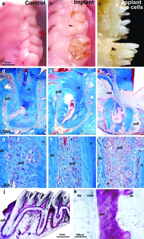
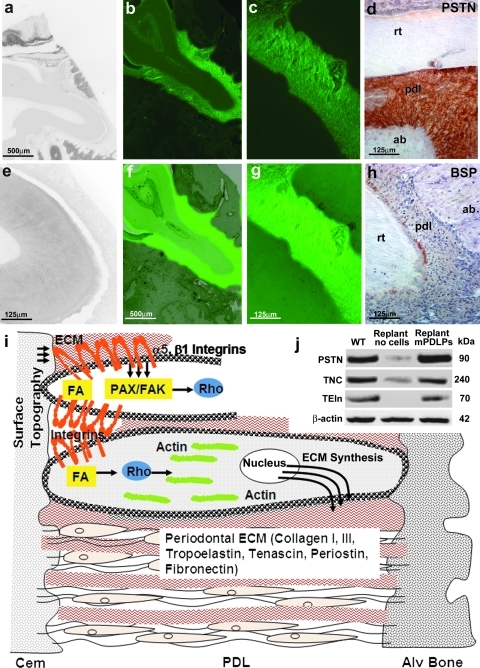
Dimension and structure of extracellular matrix surfaces have powerful influences on cell shape, adhesion, and gene expression. Here we show that natural tooth root topographies induce integrin-mediated extracellular matrix signaling cascades in tandem with cell elongation and polarization to generate physiological periodontium-like tissues. In this study we replanted surface topography instructed periodontal progenitors into rat alveolar bone sockets for 8 and 16 weeks, resulting in complete reattachment of tooth roots to the surrounding alveolar bone with a periodontal fiber apparatus closely matching physiological controls along the entire root surface. Displacement studies and biochemical analyses confirmed that progenitor-based engineered periodontal tissues were similar to control teeth and uniquely derived from preimplantation green fluorescent protein (GFP)-labeled progenitors. Together, these studies illustrate the capacity of natural extracellular surface topographies to instruct progenitor cell populations to fully regenerate complex cellular and structural morphologies of tissues once lost to disease. We suggest that our strategy could be used for the replantation of teeth lost due to trauma or as a novel approach for tooth replacement using tooth-shaped replicas.


Figures

FIG. 1.
FIG. 1.
Effect of tooth root surface topography on initial attachment and spreading of mPDLPs. Under the scanning electron microscope the natural root surface of a rat mandibular first molar exhibits an intricate, heavily grooved topography. The microporous topography of a ethylenediaminetetraacetic acid-etched root surface (a, f) had a significantly more pronounced surface relief than a nanostructured, nano-HA apatite surface (b) or a smoothened and polished root surface (d). Differences in surface topography resulted in significant changes in cell shape and attachment when apatite surfaces were seeded with mouse periodontal progenitor cells. On nano-HA, cells were flattened and spread out (c), on smoothened root surface apatite there was little or no attachment of cells (e), and on a microporous root surface, cells demonstrated an elongated, fibroblast-like morphology (g). Compared to mPDLPs grown on smooth or nano-HA surfaces, mPDLPs grown on microporous root surface apatite were significantly more elongated (h). Western blots demonstrated that the two early attachment focal adhesion proteins phospho-paxillin Y31 and phospho-focal adhesion kinase Y397 were highly expressed on cells attached to microporous root surfaces, whereas expression of these adhesion proteins was reduced in cells on nano-HA surfaces and almost absent in cells cultured on smoothened apatite surfaces (i). Changes in gene expression as a result of surface topography were not unique to native rat molar tooth roots but also occurred on smoothened or roughened apatite surfaces of identical chemical composition (j–l). (m–r) Images document the multipotency of mPLDPs to differentiate toward adipogenic (n), osteogenic (p), and chondrogenic (r) lineages compared to noninduced negative controls (m, o, q). mPDLP, mouse periodontal ligament progenitor cell; nano-HA, nano-hydroxyapatite; PAX, paxillin; FAK, focal adhesion kinase.
FIG. 2.
FIG. 2.
Attachment and growth of mPDLPs on root surfaces of extracted teeth in vitro. (b, c, d) Light microscopic images of mPDLPs attached to denuded first maxillary molars and cultured in vitro for 3 days before replantation in the tooth socket. Note the extension of fiber bundles and progenitor cells at the apical tip of the cultured implant (arrow, d). (a) A denuded rat first maxillary molar before treatment with mPDLPs. (e, f) The distribution and morphology of mPDLPs seeded on rat first maxillary molars after 10 days of culture using scanning electron microscopy. Note the PDL-like fibrous outgrowth of parallel-aligned and elongated PDL-like cells at the apical end of the tooth root. Histological analysis revealed fibrous attachment (arrow, h) of mPDLPs on root surfaces after 10 days (h) compared to untreated controls (g). Western blot (i) document significant changes in protein expression after PDL progenitors were exposed to micropatterned 3D surfaces. (i) Micropatterned 3D environments were created either by 3D cell culture in conjunction with micropatterned tooth root surfaces (center column: 3D) or after in vivo replantation for 8 weeks (right column: in vivo), and protein expression levels were compared to mPDLP expression levels in 2D culture without any microstructural challenge (left column: 2D). For all six proteins investigated (β1 integrin, α5 integrin, fibronectin, Rho A, F-actin, and periostin), protein levels were higher after exposure to 3D micropatterned root surfaces (i). Integrin α5β1 blockage (k) resulted in a loss of actin fibers and polarization when compared to control mPDLPs cultured on fibronectin coated plates (j). (l–o) Images compares various progenitor lineages and preosteoblast MC3T3 cells in response to microstructured surface topography. mPDLPs demonstrated highly elongated, fibroblast-like shaped cells aligned perpendicular to the microstructured apatite chips (l), whereas dental follicle progenitors (m), dental pulp cells (n), and MC3T3 cells (o) formed small and polygonal cells surrounding the microstructured apatite chips. rt, root; fib, fibers; GAPDH, glyceraldehyde 3-phophate dehydrogenase; PSTN, periostin.
FIG. 3.
FIG. 3.
Periodontal progenitor-driven new attachment of denuded teeth after 8 weeks of implantation in a tooth molar socket. Vertical columns: (a, d, g) WT controls; (b, e, h) replanted mPDLP-treated molars; and (c, f, i) replanted molars that were not treated with progenitor cells before replantation. Horizontal rows: (a–c) oral micrographs of rat upper right molar tooth rows; (d–f) overview histological preparations documenting the root surface/ligament interface of an entire upper first molar tooth root; and (g–i) detailed histological micrographs of the root surface/PDL/alveolar bone interface in all three groups. (b, e, h) Complete anatomical and histological integration of denuded and then mPDLP-treated rat first molars after 8 weeks of reimplantation. Reimplanted rat molars that were not subjected to progenitor cell reattachment were either lost, partially exfoliated (c), or partially resorbed (f, i). Our replantation approach resulted in the progenitor-based periodontal tissue engineering of the entire periodontium of a first maxillary molar (j, k). (j, k) Ultrathin ground sections in which the periodontium was stained with fuchsin. (j) The background outside of the fixed tooth organ was digitally removed and no other alterations were applied to the micrograph. Individual tissues are labeled for orientation. de, dentin; cem, cementum; pdl, periodontal ligament; ab, alveolar bone; res, resorption site; rt, root; m1, first maxillary rat molar. WT, wild type.
FIG. 4.
FIG. 4.
Micro-CT, scanning electron microscopic analysis, and mechanical functional test of progenitor cell-treated reimplanted teeth versus replants without progenitor cell pretreament. (a, b) 3D-reconstructed micro-CT images of replanted rat molars that were either repopulated with periodontal progenitors (a) or left untreated (b) after 16 weeks of replantation. (c–f) Higher magnification micro-CT sections (c, e) or scanning electron micrographs (d, f) of a single first molar mesial root from progenitor-treated (c, d) and untreated (e, f) replanted teeth 16 weeks postreplantation. Micrographs show optimum microanatomical integration of periodontal progenitor-treated teeth (a, c, d) in contrast to resorption, fracture, and partial ankylosis (b, e, f) in untreated controls. Individual tissues were labeled for orientation purposes. Levels of tooth displacement in response to mechanical loading (h) were very similar between progenitor-treated replants and WT controls, whereas nontreated replants were lost, loose, or ankylosed (respective percentages provided in g). CT, computed tomography; cr, crown; res, resorption; rt, root; ab, alveolar bone; de, dentin; ank, ankylosis; cem, cementum.
FIG. 5.
FIG. 5.
Molecular characterization of the attachment apparatus of replanted teeth by molecular tracing, immunohistochemisty, and Western blotting. Fluorescent micrographs illustrated green fluorescence throughout the entire newly formed PDL (b, c, f, g), whereas there was no fluorescence in nontreated replanted teeth (a, e), suggesting that the newly formed periodontium was formed by GFP-labeled, periodontal progenitors seeded on denuded root surfaces before replantation. (b, c) GFP images; (a, e and f, g) overlays of the respective GFP and phase-contrast images at a magnification of 5 × (a, b, f; scale bar = 500 μm) and 20 × (c, e, g; scale bar = 125 μm). (d, h) Immunohistostains for periostin (d) and BSP (h) on paraffin sections of mPDLP seeded first maxillary molars that were replanted into the tooth socket and maintained in vivo for 8 weeks. (d) Note the intense localization of periostin along the newly synthesized PDL fibers as seen in the native PDL. BSP expression was specifically localized at the apical root tip (h). (j) Similar expression levels for the ECM proteins PSTN, TNC, and TEIn between the progenitor cell-treated replants and WT controls as demonstrated by Western blot. In contrast, expression levels for these genes in cell-free replants were either low (periostin and tenascin C) or nondetectable (tropoelastin). (i) The sketch illustrates a simplified model of the effect of surface topography on periodontal progenitor cell shape and gene expression. Integrin surface receptors feed PDL cells with information about surrounding surfaces via the adhesome gene network. Integrin assembly and signal transduction cascades then affect intracellular machineries, including focal adhesion kinases and paxillins, which in turn regulate GTPases such as Rho to modulate actin microfilament polymerization and associated cytoskeletal changes. These changes cause PDL progenitors to elongate and stretch. In addition, intracellular integrin pathways also affect ECM gene expression, including collagens and periodontal matrix related proteins such as periostin. Thus, through the adhesome and associated integrin receptors, cell surfaces affect both periodontal cell shape and periodontal ECM gene expression, providing tissue-specific control over progenitor fate determination in the periodontal region. ECM, extracellular matrix; TNC, tenascin C; TEIn, tropoelastin; GFP, green fluorescent protein; BSP, bone sialoprotein.

Similar articles

Cited by

References

    1. Tan W. Desai T.A. Microfluidic patterning of cells in extracellular matrix biopolymers: effects of channel size, cell type, and matrix composition on pattern integrity. Tissue Eng. 2003;9:255. - PubMed
    1. Norman J.J. Desai T.A. Methods for fabrication of nanoscale topography for tissue engineering scaffolds. Ann Biomed Eng. 2006;34:89. - PubMed
    1. Traphagen S. Yelick P.C. Reclaiming a natural beauty: whole-organ engineering with natural extracellular materials. Regen Med. 2009;4:747. - PMC - PubMed
    1. Matsuura M. Herr Y. Han K.Y. Lin W.L. Genco R.J. Cho M. I Immunohistochemical expression of extracellular matrix components of normal, healing periodontal tissues in the beagle dog. J Periodontol. 1995;66:579. - PubMed
    1. Waddington R.J. Embery G. Proteoglycans and orthodontic tooth movement. J Orthod. 2001;28:281. - PubMed

Publication types

LinkOut - more resources