. 2010 Sep;35(9):799-805.
doi: 10.3109/02713683.2010.494239.

MMP-7 knock-in corneal fibroblast cell lines secrete MMP-7 with proteolytic activity towards collagen XVIII

Affiliations

MMP-7 knock-in corneal fibroblast cell lines secrete MMP-7 with proteolytic activity towards collagen XVIII

Shu-I Yeh et al. Curr Eye Res. 2010 Sep.

Abstract

Purpose: To determine whether matrix metalloproteinase-7 (MMP-7) that is stably overexpressed by mouse corneal fibroblast cell lines exhibits proteolytic activity against the NC1 fragment of collagen XVIII.

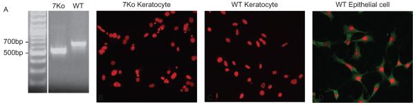
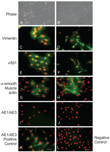
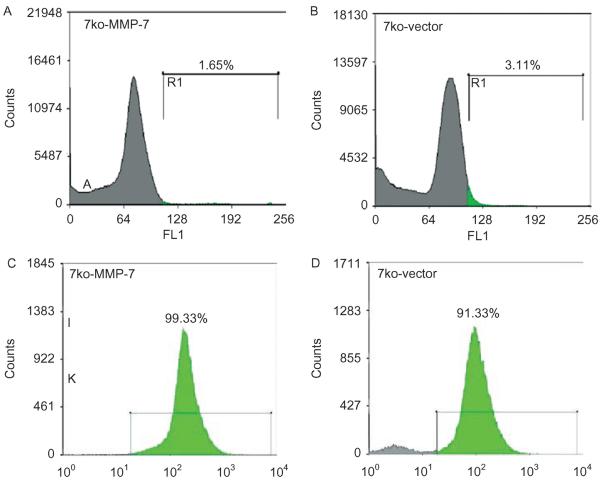
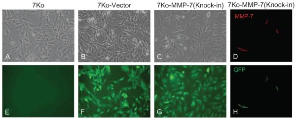
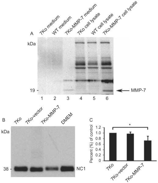
Methods: Corneal fibroblasts isolated from MMP-7 knockout (7ko) mice were subjected to SV40 T-antigen immortalization and stably transfected with a bicistronic retroviral vector encoding green fluorescence protein and active MMP-7. The resulting MMP-7 knock-in fibroblasts (7ko-MMP-7 cells) were isolated and enriched by fluorescence activated cell sorting (FACS). Culture media samples from 7ko and 7ko-MMP-7 cells were then incubated with the recombinant NC1 fragment of collagen XVIII, and NC1 degradation was monitored by immunoblotting.

Results: Immunoblot analysis revealed that MMP-7 was present in lysates and culture media from 7ko-MMP-7 fibroblasts, but not media from immortalized 7ko fibroblasts. Importantly, lower amounts of the NC1 fragment were present in in vitro enzymatic reaction mixtures containing concentrated 7ko-MMP-7 media than in those containing concentrated 7ko media.

Conclusion: Immortalized fibroblasts stably transfected with MMP-7 secrete active MMP-7 with proteolytic activity towards the NC1 fragment of collagen XVIII.


Figures

FIGURE 1
FIGURE 1
PCR analysis of the MMP-7 allele in 7ko and WT cell lines. Bands of 535 bp and 685 bp were expected for 7ko and WT cell lines, respectively (A). No immunostaining for MMP-7 was observed in WT or 7ko immortalized fibroblasts [(B) and (C); PI nuclei, red], and MMP-7 was localized to the cytoplasm of the immortalized WT corneal epithelial cells (D).
FIGURE 2
FIGURE 2
Characterization of immortalized WT and 7ko fibroblasts. WT and 7ko fibroblasts were visualized by phase contrast [WT in (A), 7ko in (B)] and immunostained for vimentin [WT in (C), 7ko in (D)], integrin α5β1 [WT in (E), 7ko in (F)], and epithelial keratin [using AE1/AE3 antibody; WT in (I), 7ko in (J)]. A subset of cells was stimulated with TGF-β and stained for α-SMA (myofibroblast marker) [WT in (G), 7ko in (H)]. Corneal epithelial cells immunostained for epithelial keratin served as a positive control (K), and TGF-β-stimulated corneal epithelial cells immunostained for α-SMA served as a negative control (L). Scale bar, 50 μm.
FIGURE 3
FIGURE 3
Enrichment of GFP-expressing cells with FACS. Initial sorting yielded only 1.65% GFP-expressing 7ko-MMP-7 fibroblasts (A) and 3.11% 7ko-vector fibroblasts (B). After three sequential sortings, the population of GFP-expressing cells exceeded 90% purity for both the 7ko-MMP-7 (C) and 7ko-vector group (D).
FIGURE 4
FIGURE 4
GFP and MMP-7 expression in cultured fibroblasts. Phase contrast images of 7ko (A), 7ko-vector (B), and 7ko-MMP-7 knock-in fibroblasts (C). No GFP expression was observed in 7ko fibroblasts (E). Confocal images of 7ko-vector (F) and 7ko-MMP-7 (knock-in) fibroblasts [(G) and (H)]. There was detectable MMP-7 immunostaining in 7ko-MMP-7 knock-in fibroblasts (D).
FIGURE 5
FIGURE 5
Immunoblot analysis of MMP-7 expression in media (lanes 1–3) and lysates (lanes 4–6) from WT, 7ko, and 7ko-MMP-7 fibroblasts (A). The 7ko and WT groups served as negative controls. Immunoblot analysis of collagen XVIII NC1 degradation induced by media collected from 7ko, 7ko-vector, and 7ko-MMP-7 fibroblast cultures (B). Three independent experiments were performed to quantify the intensity of remaining, unprocessed NC1 fragments. The NC1 fragment density in the 7ko-MMP-7 group is 0.72 ± 0.15% of that of the control 7ko group (C).

Similar articles

Cited by

References

    1. Ellenberg D, Azar DT, Hallak JA, et al. Novel aspects of corneal angiogenic and lymphangiogenic privilege. Prog Retin Eye Res. 2010;29:208–248. - PMC - PubMed
    1. Azar DT. Corneal angiogenic privilege: Angiogenic and antiangiogenic factors in corneal avascularity, vasculogenesis, and wound healing (an American Ophthalmological Society thesis) Trans Am Ophthalmol Soc. 2006;104:264–302. - PMC - PubMed
    1. Ambati BK, Nozaki M, Singh N, et al. Corneal avascularity is due to soluble VEGF receptor-1. Nature. 2006;443:993–997. - PMC - PubMed
    1. Albuquerque RJ, Hayashi T, Cho WG, et al. Alternatively spliced vascular endothelial growth factor receptor-2 is an essential endogenous inhibitor of lymphatic vessel growth. Nat Med. 2009;15:1023–1030. - PMC - PubMed
    1. Daniels JT, Cambrey AD, Occleston NL, et al. Matrix metalloproteinase inhibition modulates fibroblast-mediated matrix contraction and collagen production in vitro. Invest Ophthalmol Vis Sci. 2003;44:1104–1110. - PubMed

Publication types

Substances

LinkOut - more resources