Case Reports
. 2010 Aug 26:8:74.
doi: 10.1186/1477-7819-8-74.

Adenoid cystic carcinoma of the peripheral lung: a case report

Affiliations
Case Reports

Adenoid cystic carcinoma of the peripheral lung: a case report

Masahiro Kitada et al. World J Surg Oncol. .

Abstract

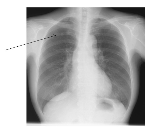
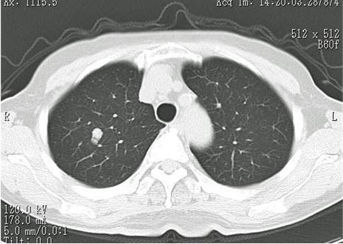
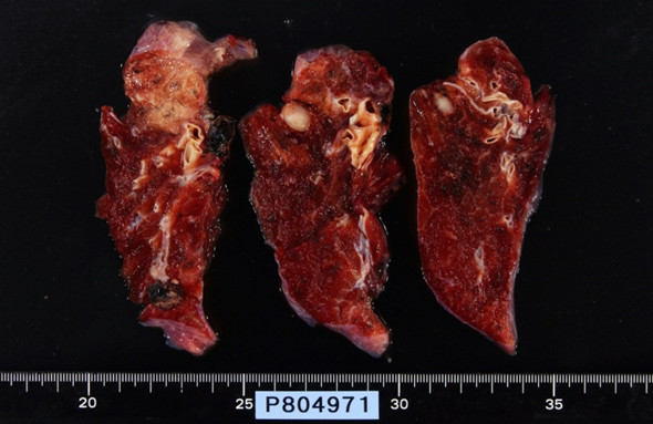
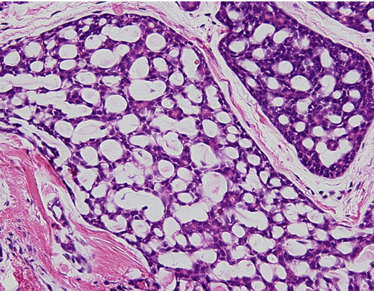
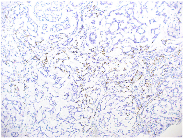
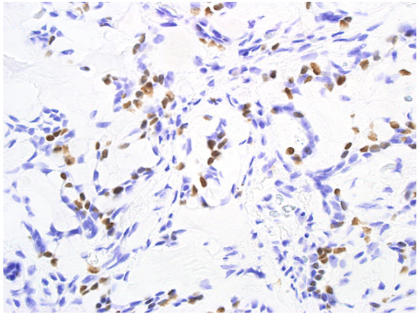
Adenoid cystic carcinoma of the peripheral lung is a rare entity. We recently encountered a patient with adenoid cystic carcinoma. A 75-year-old woman showed a nodular lesion with 10 mm in diameter in the right upper lung field on chest radiography. The diagnosis was unclear, but lung cancer could not be ruled out. Thoracoscopic biopsy was performed, and intraoperative pathological diagnosis revealed the carcinoma of the lung. We enforced upper lobectomy and mediastinal lymph node dissection to the patient. Histopathological examination revealed adenoid cystic carcinoma with a characteristic cribriform structure. Immunohistochemical examination revealed that the tumor cells were positive for thyroid transcription factor 1 (TTF-1), this tumor was diagnosed primary ACC of the lung.


Figures

Figure 1
Figure 1
Chest radiography showing a nodular shadow in the upper lung field.
Figure 2
Figure 2
Chest CT showing the tumor, 10 mm in diameter, in S1.
Figure 3
Figure 3
Macroscopic findings showing a tumor with clear boundaries, a uniform 10 mm in diameter.
Figure 4
Figure 4
Histological findings showing typical cribriform pattern and constituent cells including of bronchial epithelial cells, myoepithelial cells, and basal cells (HE ×400).
Figure 5
Figure 5
Immunohistochemical examination revealed that tumor cells were positive for TTF-1 (×100).
Figure 6
Figure 6
Immunohistochemical examination revealed that tumor cells were positive for TTF-1(×400).

References

    1. Travis WD, Travis LB, Devesa SS. Lung cancer. Cancer. 1995;75:191–02. doi: 10.1002/1097-0142(19950101)75:1+<191::AID-CNCR2820751307>3.0.CO;2-Y. - DOI - PubMed
    1. Kawashima O, Hirai T, Kamyoshihara M, Ishikawa S, Morishita Y. Primary adenoid cystic carcinoma of the lung: report of two cases and therapeuticconsiderations. Lung Cancer. 1988;19:211–7. doi: 10.1016/S0169-5002(97)00098-6. - DOI - PubMed
    1. Inoue H, Iwashita A, Kanegae H, Higuchi K, Fujinaga Y, Matsumoto I. Peripheral pulmonary adenoid cystic carcinoma with substantial submucosal extension to the proximal bronchus. Thorax. 1991;46:147–8. doi: 10.1136/thx.46.2.147. - DOI - PMC - PubMed
    1. Berghmans T, Paesmans M, Mascaux C, Martin B, Meert AP, Haller A, Lafitte JJ, Sculier JP. Thyroid transcription factor 1--a new prognostic factor in lung cancer: a meta-analysis. Ann Oncol. 2006;16:1673–6. doi: 10.1093/annonc/mdl287. - DOI - PubMed
    1. Jagirdar J. Application of immunohistochemistry to the diagnosis of primary and metastatic carcinoma to the lung. Arch pathol Lab Med. 2008;132:386–96. - PubMed

Publication types