Review
. 2011 May;29(3):295-304.
doi: 10.1016/j.ijdevneu.2010.08.003. Epub 2010 Aug 24.

Insights into the neurodevelopmental origin of schizophrenia from postmortem studies of prefrontal cortical circuitry

Affiliations
Review

Insights into the neurodevelopmental origin of schizophrenia from postmortem studies of prefrontal cortical circuitry

Monica Beneyto et al. Int J Dev Neurosci. 2011 May.

Abstract

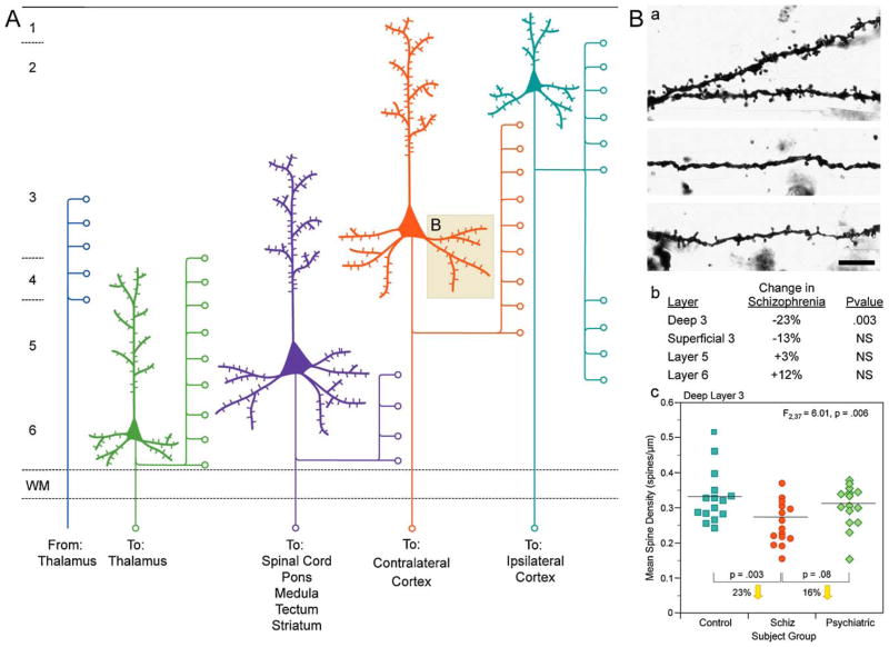
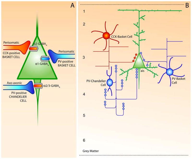
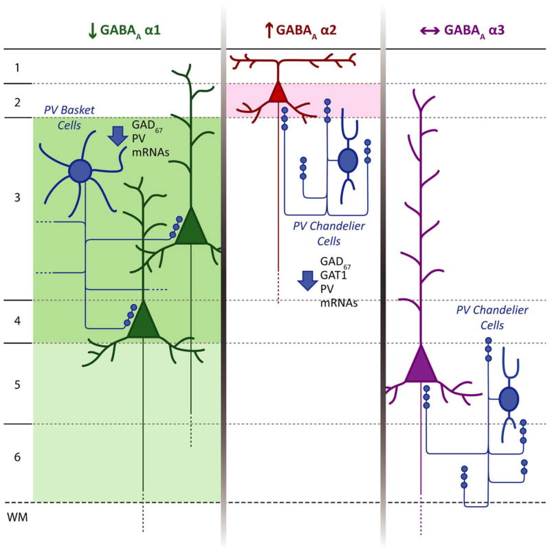
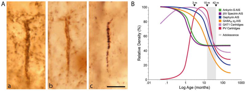
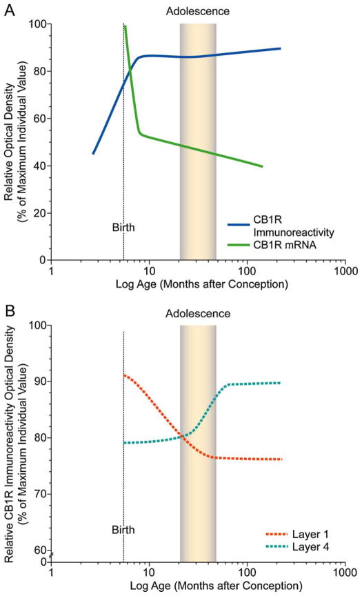
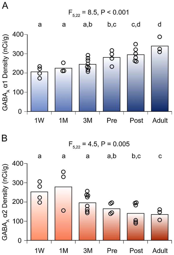
The hypothesis that schizophrenia results from a developmental, as opposed to a degenerative, process affecting the connectivity and network plasticity of the cerebral cortex is supported by findings from morphological and molecular postmortem studies. Specifically, abnormalities in the expression of protein markers of GABA neurotransmission and the lamina- and circuit-specificity of these changes in the cortex in schizophrenia, in concert with knowledge of their developmental trajectories, offer crucial insight into the vulnerability of specific cortical networks to environmental insults during different periods of development. These findings reveal potential targets for therapeutic interventions to improve cognitive function in individuals with schizophrenia, and provide guidance for future preventive strategies to preserve cortical neurotransmission in at-risk individuals.


Figures

Fig. 1
Fig. 1
(A) Schematic representation of the main afferent and efferent pyramidal connections to and from specific cortical lamina. (B) Reduction in pyramidal neuron dendritic spines in deep layer 3 of the DLPFC in schizophrenia. (a) Golgi-impregnated basilar dendrites and spines on deep layer 3 pyramidal neurons from a normal comparison (top) and two subjects with schizophrenia (bottom). Note the reduced density of spines in the subjects with schizophrenia in these extreme examples. (b) Laminar specificity of the spine density differences in the subjects with schizophrenia relative to normal control subjects. (c) Scatter plot demonstrating the lower density of spines on the basilar dendrites of deep layer 3 pyramidal neurons in the DLPFC of subjects with schizophrenia relative to both normal and psychiatrically ill comparison subjects. Adapted from Lewis and Gonzalez-Burgos (2008).
Fig. 2
Fig. 2
(A) Schematic summary of cortical GABA circuits describing the subcellular location and subunit-specificity of the receptors targeted by different GABA neuron subtypes. (B) Morphological and biochemical features of PV- and CCK-positive subpopulations of cortical GABA neurons in the DLPFC. The chandelier and basket neurons provide inhibitory input to the axon initial segment (ais) and the cell body and proximal dendrites, respectively, of pyramidal neurons. 1–4, layers of DLPFC. Adapted from Lewis et al. (2005).
Fig. 3
Fig. 3
Schematic summary of hypothesized circuit-specific transcript alterations in pre- and postsynaptic markers of GABA neurotransmission in the DLPFC of subjects with schizophrenia. For each GABAA α subunit, the background shading marks the cortical layers where the indicated change in expression of that subunit was found. The laminar specificity of the decrease in α1 expression matches that of the alterations in GAD67 and PV mRNAs thought to be present in PV+ basket cells. The increase in α2 expression in layer 2 is consistent with previous findings of pre- and postsynaptic alterations in chandelier cell inputs to the axon initial segment of pyramidal cells in this location. In contrast, the absence of alterations in α3 subunit expression, which is present postsynaptic to chandelier cells in deep layer pyramidal neurons matches the failure to find significant changes in chandelier cell inputs in these layers. Adapted from Beneyto et al. (2010).
Fig. 4
Fig. 4
(A) Pre- and postsynaptic markers of chandelier neuron inputs to the axon initial segment of pyramidal neurons. (a and b) Immunoreactivity for GAT1 (a) and parvalbumin (b) clearly identifies vertical arrays of chandelier neuron axon terminals (cartridges) that are located below the cell bodies of unlabelled pyramidal neurons. (c) Immunoreactivity for the GABAA α2 subunit is localized postsynaptically, in the axon initial segment of pyramidal neurons. From Lewis et al. (2005). (B) Schematic summary of the trajectories of pyramidal neuron axon initial segment and chandelier neuron axon cartridges labeled with different markers across postnatal development in area 46 of monkey DLPFC. Lines for each marker represent the percent maximal value achieved plotted against age in months after birth on a log scale. Arrowheads demarcate the indicated ages in months, and the shaded area indicates the approximate age range corresponding to adolescence in this species. From Cruz et al. (2009a,b).
Fig. 5
Fig. 5
Schematic summary figures illustrating trajectories of overall CB1R immunoreactivity and mRNA levels (A) and density of CB1R-IR axons in layers 1 and 4 (B) across development of monkey DLPFC. Lines were generated by plotting the density as a percent of the maximum density value for individual animals for each marker as a function of age in months after conception on a log scale, fitted by Loess regression analysis, and smoothed by hand. The shaded area indicates the approximate age range corresponding to adolescence (15–42 months; Plant 1988) in macaque monkey. Note the different developmental time courses in overall CB1R immunoreactivity and mRNA levels (A) and in the laminar distribution of CB1R immunoreactivity (B). From Eggan et al. (2010).
Fig. 6
Fig. 6
Expression levels of GABAA receptor α1 and α2 subunit mRNAs in the monkey DLPFC during development. The optical densities for α1 (A) and α2 (B) subunit mRNAs within area 46 are individually plotted for each age group. The mean values for each age group are indicated as bars. During postnatal development, α1 mRNA levels increased, whereas α2 mRNA levels declined. Age groups that do not share the same letter are statistically different at α ≤ 05. Note that the shift in α subunit expression is a progressive and protracted process that lasts through adolescence. M, months; Pre, prepuberty; Post, postpuberty; W, week. From Hashimoto et al. (2009).

Similar articles

Cited by

References

    1. Akbarian S, Huang HS. Molecular and cellular mechanisms of altered GAD1/GAD67 expression in schizophrenia and related disorders. Brain Res Rev. 2006;52:293–304. - PubMed
    1. Akbarian S, Huntsman MM, Kim JJ, Tafazzoli A, Potkin SG, Bunney WE, Jr, Jones EG. GABAA receptor subunit gene expression in human prefrontal cortex: comparison of schizophrenics and controls. Cereb Cortex. 1995a;5:550–560. - PubMed
    1. Akbarian S, Kim JJ, Potkin SG, Hagman JO, Tafazzoli A, Bunney WE, Jr, Jones EG. Gene expression for glutamic acid decarboxylase is reduced without loss of neurons in prefrontal cortex of schizophrenics. Arch Gen Psychiatry. 1995b;52:258–266. - PubMed
    1. Alexander GE, Goldman PS. Functional development of the dorsolateral prefrontal cortex: an analysis utlizing reversible cryogenic depression. Brain Res. 1978;143:233–249. - PubMed
    1. Anderson SA, Classey JD, Conde F, Lund JS, Lewis DA. Synchronous development of pyramidal neuron dendritic spines and parvalbumin-immunoreactive chandelier neuron axon terminals in layer III of monkey prefrontal cortex. Neuroscience. 1995;67:7–22. - PubMed

MeSH terms