. 2010 Aug 27;17(8):892-902.
doi: 10.1016/j.chembiol.2010.06.006.

Modulation of pantothenate kinase 3 activity by small molecules that interact with the substrate/allosteric regulatory domain

Affiliations

Modulation of pantothenate kinase 3 activity by small molecules that interact with the substrate/allosteric regulatory domain

Roberta Leonardi et al. Chem Biol. .

Abstract

Pantothenate kinase (PanK) catalyzes the rate-controlling step in coenzyme A (CoA) biosynthesis. PanK3 is stringently regulated by acetyl-CoA and uses an ordered kinetic mechanism with ATP as the leading substrate. Biochemical analysis of site-directed mutants indicates that pantothenate binds in a tunnel adjacent to the active site that is occupied by the pantothenate moiety of the acetyl-CoA regulator in the PanK3acetyl-CoA binary complex. A high-throughput screen for PanK3 inhibitors and activators was applied to a bioactive compound library. Thiazolidinediones, sulfonylureas and steroids were inhibitors, and fatty acyl-amides and tamoxifen were activators. The PanK3 activators and inhibitors either stimulated or repressed CoA biosynthesis in HepG2/C3A cells. The flexible allosteric acetyl-CoA regulatory domain of PanK3 also binds the substrates, pantothenate and pantetheine, and small molecule inhibitors and activators to modulate PanK3 activity.


Figures

FIGURE 1
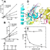
FIGURE 1. Biochemical mechanism and structure of PanK3
Graphical analysis of product inhibition experiments using fixed concentrations of ADP and variable concentrations of either ATP (A) or pantothenate (B) showed that ADP was competitive with ATP and uncompetitive with pantothenate. (C) These data are consistent with a compulsory ordered kinetic mechanism with ATP as the leading substrate and ADP as the last product to leave the enzyme. (D) Detailed interactions of the pantothenate portion of CoA within the tunnel formed from the stable β10 and β11 platform of the ATP-binding domain (purple) on one subunit (yellow) and the flexible loop between α7′ and β13′ that reaches over from the opposite monomer (cyan). The residues investigated by site-directed mutagenesis are shown. Acetyl-CoA is shown as a ball-and–stick representation (grey carbons) and the P-loop is colored magenta. Red spheres are water molecules and hydrogen bonds are indicated by dotted red lines. Note that in this ‘open’ conformation, the general base E138 is not positioned to perform catalysis and the adenine ring of acetyl-CoA acts as a wedge to prevent domain closure. See Supplementary Experimental Procedures for structure determination and Table 1 for the structural statistics. (E) PanK3(S195V) was refractory to acetyl-CoA inhibition compared to PanK3. PanK3 activity in the absence of acetyl-CoA was set at 100%. PanK3(S195V) has a KM defect for pantothenate (see text). The data were duplicate measurements with the standard error indicated by the bars.
FIGURE 2
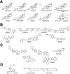
FIGURE 2. Structures of the biochemically characterized inhibitors and activators identified in the HTS
(A) Steroids and fusidic acid. (B) Thiazolidedione and non-thiazolidinedione PPARγ ligands. (C) Sulfonylurea and non sulfonylurea insulin secretagogues. (D) PanK3 activators.
FIGURE 3
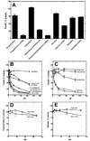
FIGURE 3. PanK3 inhibition by selected compounds identified by HTS
The PanK3 radioactive assay described under “Experimental Procedures” was used to test selected HTS hits and related molecules. (A) Steroids and their sulfate derivatives were tested at 100 µM concentration against PanK3. (B) The thiazolidinedione PPARγ ligands evaluated were rosiglitazone (●), pioglitazone (○), ciglitazone (■) and MCC-555 (□). Three structurally unrelated PPARγ ligands GW501516 (△), GW1929 (◆) and GW9662 (▲) were also assayed. (C) Sulfonylurea insulin secretagogues identified in the HTS, glyburide (●) and glipizide (○) were validated PanK3 inhibitors. Three other insulin secretagogues analyzed were tolbutamide (□), repaglinide (■) and nateglinide (◆). PanK3 activity in the presence of DMSO only was set at 100%. (D) PanK3(S195V) was refractory to inhibition by MCC-555 (□) and glyburide (■). (E) SaPanK was not inhibited by either MCC-555 (□) or glyburide (■). PanK3 activity in the presence of DMSO only was set at 100%. The specific activities used as 100% in these assays were: PanK3, 149 ± 2 nmoles/min/mg; PanK3(S195V), 48 ± 3 nmoles/min/mg. The data were duplicate measurements with the standard error indicated by the bars. IC50 values ± standard error were derived from fitting the 14-point dataset to a single-site binding model using GraphPad software. See Supplementary Fig. S2 for the kinetic analysis of MCC-555 and glyburide inhibition of PanK3 and Supplementary Fig. S3 for the isoform selectivity of MCC-555, glyburide and tamoxifen.
FIGURE 4
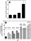
FIGURE 4. The thiazolidinediones inhibit CoA biosynthesis in cell culture
(A) Total RNA was extracted from HepG2/C3A cells and real-time PCR was used to determine the relative abundance of the PanK isoform mRNA present normalized to GAPDH as the calibrator. (B) HepG2/C3A cells were labeled with [3H]pantothenate and the amount of label incorporated into CoA was determined as described under Experimental Procedures. The amount of incorporation in the control experiment in the presence of DMSO was set at 100%, and the incorporation rates in the presence of the test compounds was expressed as a percent of this value. Hopantenate (Hopan), a known inhibitor of CoA biosynthesis (Zhang et al., 2007) was used as a positive control, MCC-555, rosiglitazone (Rosiglit.), glyburide, oleoyl-carnitine, oleoyl-ethanolaminde and tamoxifen were tested at the indicated concentrations. The data were duplicate measurements with the standard error indicated by the bars.
FIGURE 5
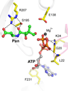
FIGURE 5. Model of the PanK3 ‘closed’ active site
The model is based on the known structure of the homologous SaPanK closed active site. Pantothenate (Pan) carbons are green, protein residues are yellow, Mg2+ is a dark grey ball, and ATP carbons are grey. Predicted hydrogen bonds are shown as dashed black lines. The protein backbone is transparent grey, and the P-loop is rendered as transparent magenta. Note that Glu138 is well positioned to act as a general base, unlike the position shown in Fig. 1D. Also, the phosphate groups of ATP engage the P-loop and the adenine ring occupies a pocket that is distinct from that which binds the adenine ring of acetyl-CoA (Fig. 1D).

References

    1. Bell DS. Practical considerations and guidelines for dosing sulfonylureas as monotherapy or combination therapy. Clin. Ther. 2004;26:1714–1727. - PubMed
    1. Bode KA, Donner MG, Leier I, Keppler D. Inhibition of transport across the hepatocyte canalicular membrane by the antibiotic fusidate. Biochem. Pharmacol. 2002;64:151–158. - PubMed
    1. Boyd AE, Aguilar-Bryan L, Nelson DA. Molecular mechanisms of action of glyburide on the beta cell. Am. J. Med. 1990;89:3S–10S. - PubMed
    1. Gale EA. Lessons from the glitazones: a story of drug development. Lancet. 2001;357:1870–1875. - PubMed
    1. Gibbs TT, Russek SJ, Farb DH. Sulfated steroids as endogenous neuromodulators. Pharmacol. Biochem. Behav. 2006;84:555–567. - PubMed

Publication types

MeSH terms

Associated data