. 2010 Aug 27;39(4):535-47.
doi: 10.1016/j.molcel.2010.08.004.

Identification of regulators of chaperone-mediated autophagy

Affiliations

Identification of regulators of chaperone-mediated autophagy

Urmi Bandyopadhyay et al. Mol Cell. .

Abstract

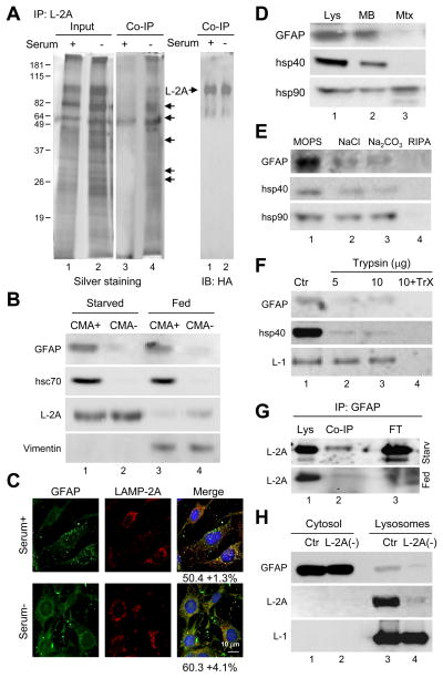
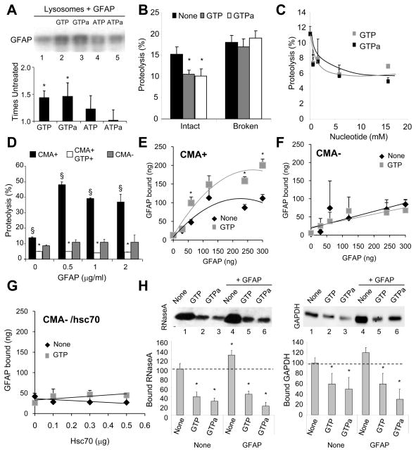
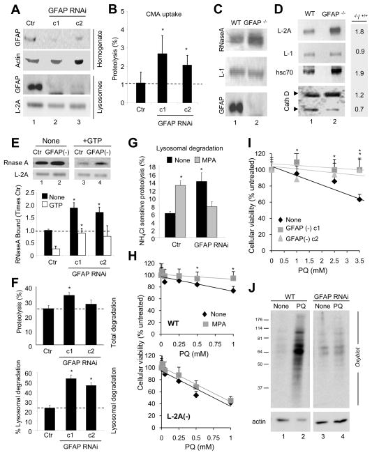
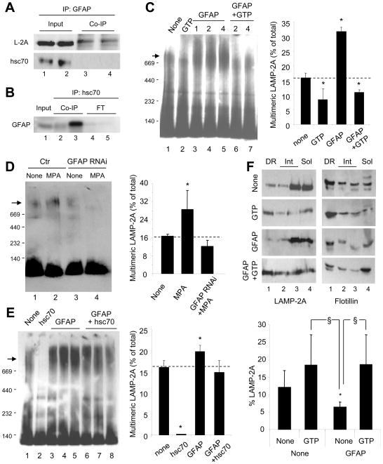
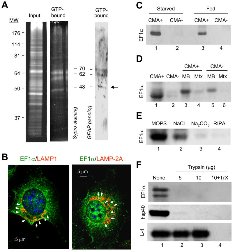
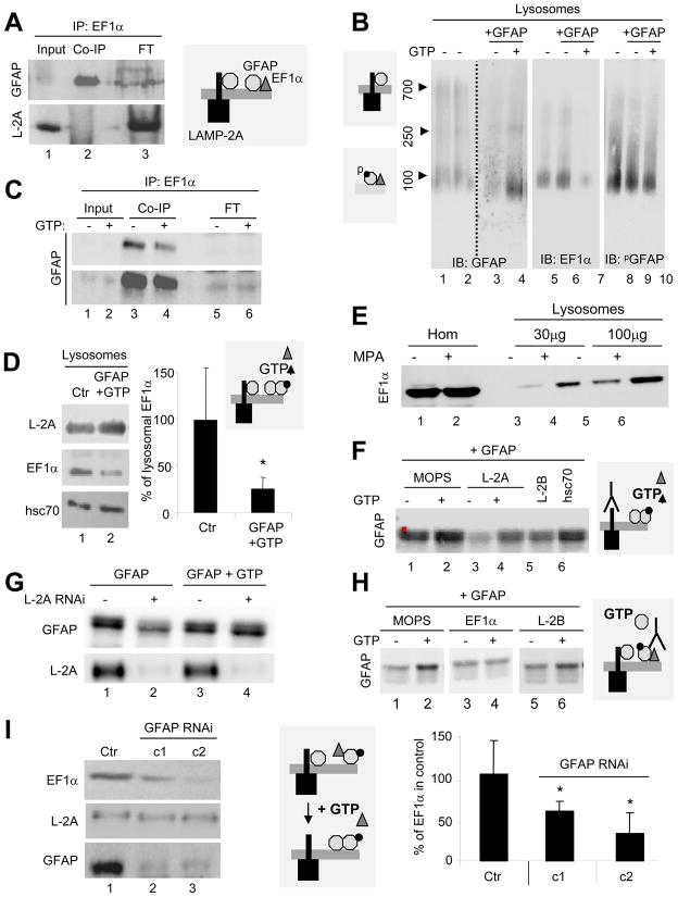
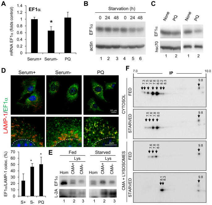
Chaperone-mediated autophagy (CMA) is a selective mechanism for the degradation of cytosolic proteins in lysosomes that contributes to cellular quality control and becomes an additional source of amino acids when nutrients are scarce. A chaperone complex delivers CMA substrates to a receptor protein at the lysosomal membrane that assembles into multimeric translocation complexes. However, the mechanisms regulating this process remain, for the most part, unknown. In this work, we have identified two regulatory proteins, GFAP and EF1alpha, that mediate a previously unknown inhibitory effect of GTP on CMA. GFAP stabilizes the multimeric translocation complex against chaperone-mediated disassembly, whereas GTP-mediated release of EF1alpha from the lysosomal membrane promotes self-association of GFAP, disassembly of the CMA translocation complex, and the consequent decrease in CMA. The dynamic interactions of these two proteins at the lysosomal membrane unveil now a role for GTP as a negative regulator of CMA.


Figures

Figure 1
Figure 1. GFAP interacts with LAMP-2A in CMA-active lysosomes
(A) Lysosomes from mouse fibroblasts expressing HA-LAMP-2A and maintained with or without serum (20 h) were incubated with an excess of RNase A and subjected to co-immunoprecipitation (Co-IP) for HA. Silver staining (left) and immunoblot for HA (right). Arrows: bands detectable by silver staining. (B) Immunoblot for the indicated proteins of liver lysosomes active (CMA+) and inactive (CMA-) for CMA from fed and 48 h starved rats. Percentages of cellular protein recovered in CMA+ fed liver lysosomes: 1% GFAP, 0.65% hsc70 and 1.7% vimentin. (C) Immunofluorescence for GFAP and LAMP-2A of mouse fibroblasts. Percentage of colocalization calculated in 20 cells is indicated. (D–F) Immunoblot for the indicated proteins of lysosomes from 48h starved rat liver (Lys) fractionated into membranes (MB) and matrices (Mtx) (D), washed with buffers of increasing stringency (E) or treated with trypsin with or without Triton X-100 (TrX) (F). (G) Co-IP for GFAP and immunoblot for LAMP-2A of lysosomes from fed or 48h starved rat livers. FT: flow through. (H) GFAP in lysosomes from control (CTR) or LAMP-2A RNAi mouse fibroblasts (L2A (-)). LAMP-1 and LAMP-2A are shown as lysosomal markers.
Figure 2
Figure 2. GFAP modulates CMA activity in a GTP-dependent manner
(A) Binding of GFAP to intact rat liver lysosomes. GTPa and ATPa: non-hydrolyzable analogues. Bottom: densitometric analysis of immunoblots (n= 5). (B) Degradation of a pool of radiolabeled cytosolic proteins by intact or broken rat liver lysosomes alone or with 1mM nucleotides (n= 4). (C) Effect of increasing concentrations of the indicated nucleotides on the proteolysis of a pool of cytosolic proteins by intact rat liver lysosomes (n= 3). (D) Effect of GFAP and/or GTP (1mM) on the proteolysis of the pool of cytosolic proteins by lysosomes active (CMA+) or inactive (CMA-) for CMA (n= 4). (E–F) Binding of GFAP to the same lysosomes as in D. (n= 4–5). (G) Effect of hsc70 and/or GTP (1mM) on the binding of GFAP (120 ng) to CMA incompetent lysosomes (n =3). (H) Binding of RNase A (left) and GAPDH (right) to intact lysosomes incubated with GTP, GTPa and GFAP, as labeled. Values are as percentage of protein bound without additions (None) (n = 6). All values are mean +S.E. p < 0.05 with control samples (*) or for GFAP samples with or without GTP (§).
Figure 3
Figure 3. Changes in CMA in cells deficient for GFAP
(A) Immunobloted for the indicated proteins in fractions from RALA cells control (Ctr) or stably RNAi for GFAP (clone 1 and 2 are against two different regions). (B) Degradation of a pool of radiolabeled cytosolic proteins by intact lysosomes show in (A). Proteolysis in control was given an arbitrary value of 1(n = 4). (C) Binding of RNase A to lysosomes from wild-type (WT) and GFAP knock-out mice (GFAP−/−). L-1: LAMP-1. (D) Immunoblot of the same lysosomes described in (C) for the indicated proteins. Right: folds-change in GFAP−/− mice compared to control. (E) Binding of RNase A to lysosomes isolated from control (Ctr) and GFAP RNAi mouse fibroblasts without (none) or with GTP (1mM). Bottom: densitometric analysis. Binding in untreated control lysosomes was given an arbitrary value of 1 (n= 4). (F) Degradation of long-lived proteins in control (Ctr) or GFAP RNAi cells. Top: Total protein degradation. Bottom: Lysosomal degradation (sensitive to ammonium chloride) (n =4). (G) Percentage of lysosomal degradation in control (Ctr) and GFAP RNAi cells untreated (None) or not with mycophenolic acid (MPA) (n= 3). (H) Viability of wild-type (WT) (top) or RNAi LAMP-2A cells (L2A(-)) (bottom) untreated (None) or treated with MPA after exposure to the indicated concentrations of paraquat (PQ) (n =4). (I) Viability of control (Ctr) and GFAP RNAi cells upon exposure to PQ (n =3). (J) Oxiblot of cells in I. All values are mean +S.E. * p<0.05 with control samples.
Figure 4
Figure 4. GFAP modifies the dynamics of the CMA receptor at the lysosomal membrane
(AB) Co-immunoprecipitation with GFAP (duplicate) (A) or with two different antibodies against hsc70 (lanes 2 and 3) (B) of lysosomes from 48h starved rat livers. (C) Blue native electrophoresis (BNE) and LAMP-2A immunoblot of rat liver lysosomes incubated alone (None) and/or with 1 mM GTP and GFAP at the indicated concentrations (mg/ml). Arrow: 700kDa translocation complex. Right: Percentage of LAMP-2A in the 700kDa complex (n= 4). (D–E) BNE for LAMP-2A of lysosomes from control (Ctr) or GFAP RNAi cells (D) or from rat liver (E) after the indicated treatments. Right: Percentage of LAMP-2A in the 700kDa complex (n= 3). (F) Immunoblot for LAMP-2A and flotillin of rat liver lysosomes incubated alone (None) or with GFAP and/or GTP and subjected to Triton X-114 extraction and sucrose density gradient floatation. Detergent-resistant (DR), intermediate (Int) or detergent-soluble (Sol) fractions are shown. Bottom: Percentage of LAMP-2A in DR (n= 5). All values are mean +S.E. p<0.05 with untreated (*) or with GTP treated (§).
Figure 5
Figure 5. The GTP-binding protein EF1α interacts with GFAP at the lysosomal membrane
(A) GTP-affinity chromatography of 48h starved rat liver lysosomal membranes. Left: SyproRuby staining. Right: Panning for GFAP of the eluted fraction. Arrow: elongation factor 1α (EF1α). (B) Immunofluorescence for LAMP-1 (left) or LAMP-2A (right) and EF1α in mouse fibroblasts maintained in the absence of serum. (C–D) Immunoblot for EF1α of starved rat liver lysosomes with high (CMA+) and low (CMA-) CMA activity (C) and their corresponding membranes (MB) and matrices (Mtx) (D). (E–F) Immunoblot of CMA active lysosomes washed with buffers of increasing stringency (E) or incubated with increasing concentrations of trypsin in presence or absence of Triton X-100 (TrX) (F).
Figure 6
Figure 6. The dynamic interaction of EF1α, GFAP and LAMP-2A at the lysosomal membrane is modulated by GTP
(A) Co-immunoprecipitation for EF1α of starved rat liver lysosomes. FT: flow through. (B) Blue native electrophoresis and immunoblot for the indicated proteins of lysosomes as in (A) incubated or not with 1mM GTP. Left: Molecular weights. (C) CO-IP for EF1α of the same lysosomes as in (B). Bottom: higher exposure. FT: flow through. (D) Immunoblot of rat liver lysosomes untreated (Ctr) or incubated with GTP and GFAP. Right: Percentage of EF1α remaining after the treatments (n= 3). (E) Immunoblot for EF1α of cell homogenates (Hom) and lysosomes from cells treated or not with mycophenolic acid (MPA). (F) Immunoblot for GFAP of rat liver lysosomes were pre-incubated in MOPS buffer alone or with antibodies against LAMP-2A (L-2A), LAMP-2B (L-2B) or hsc70, followed by GFAP with or without 1mM GTP. (G) Immunoblot of lysosomes from control or LAMP-2A RNAi cells incubated with GFAP with or without 1mM GTP. (H) Immunoblot for GFAP of rat liver lysosomes pre-incubated alone (MOPS) or with antibodies against EF1α or LAMP-2B (L-2B), followed by GFAP. (I) Immunoblot of lysosomes from control and GFAP RNAi cells (two clones shown). Right: EF1α at the lysosomal as percentage of EF1α in control (n = 3). Values are all mean +S.E. *p<0.05.
Figure 7
Figure 7. Regulation of the association of EF1α with lysosomes
(A) EF1α mRNA levels in fibroblasts maintained in the presence or absence (48h) of serum or treated with paraquat (PQ) for 6 h. Values are fold control after normalization for actin (n= 3–4). (B–C) Immunoblot of fibroblasts maintained in the absence of serum for the indicated times (B) or of livers of rats untreated (None) or treated with PQ (C). Two different sets are shown. (D) Immunofluorescence of fibroblasts maintained in presence (Serum+) or absence of serum (Serum-) for 24h or treated with PQ for 6h. Bottom: Higher magnification insets and quantification of colocalization in >25 cells (n=2). (E) Immunoblot of fed or starved (48h) rat liver homogenates (Hom) and lysosomes (Lys) active (CMA+) or inactive (CMA-) for CMA. (F) Bidimensional electrophoresis and immunoblot for EF1α of cytosol or CMA+ lysosomes isolated from fed or 48h starved rats. Arrows: Isoelectric point values.

Comment in

  • GTP: gatekeeper for autophagy.
    Beau I, Codogno P. Beau I, et al. Mol Cell. 2010 Aug 27;39(4):485-6. doi: 10.1016/j.molcel.2010.08.005. Mol Cell. 2010. PMID: 20797620

Similar articles

Cited by

References

    1. Bandyopadhyay U, Kaushik S, Vartikovsky L, Cuervo AM. Dynamic organization of the receptor for chaperone-mediated autophagy at the lysosomal membrane. Mol Cell Biol. 2008;28:5747–5763. - PMC - PubMed
    1. Boran MS, Garcia A. The cyclic GMP-protein kinase G pathway regulates cytoskeleton dynamics and motility in astrocytes. J Neurochem. 2007;102:216–230. - PubMed
    1. Coppard NJ, Clark BF, Cramer F. Methylation of elongation factor 1 alpha in mouse 3T3B and 3T3B/SV40 cells. FEBS Lett. 1983;164:330–334. - PubMed
    1. Cuervo A, Dice J. A receptor for the selective uptake and degradation of proteins by lysosomes. Science. 1996;273:501–503. - PubMed
    1. Cuervo A, Dice J. Unique properties of lamp2a compared to other lamp2 isoforms. J Cell Sci. 2000;113:4441–4450. - PubMed

Publication types

MeSH terms

Substances