. 2010 Aug 27;39(4):618-31.
doi: 10.1016/j.molcel.2010.07.025.

A proteomic screen for nucleolar SUMO targets shows SUMOylation modulates the function of Nop5/Nop58

Affiliations

A proteomic screen for nucleolar SUMO targets shows SUMOylation modulates the function of Nop5/Nop58

Belinda J Westman et al. Mol Cell. .

Abstract

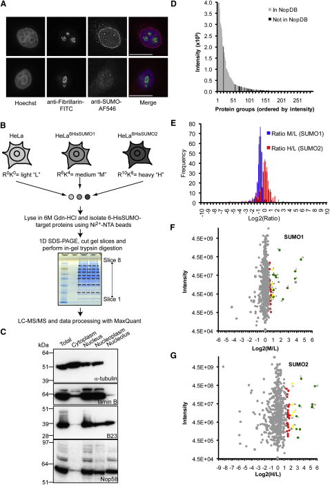
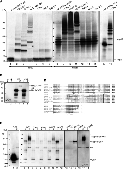
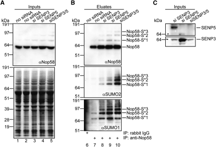
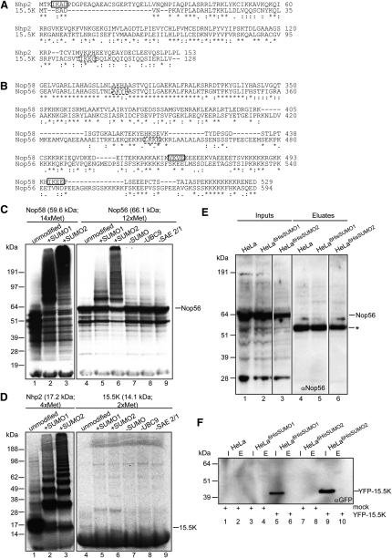
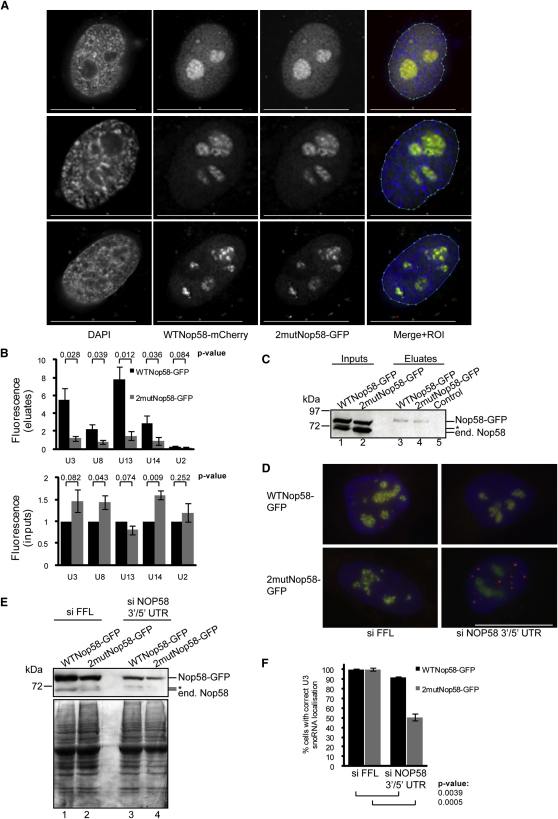
Posttranslational SUMO modification is an important mechanism of regulating protein function, especially in the cell nucleus. The nucleolus is the subnuclear organelle responsible for rRNA synthesis, processing, and assembly of the large and small ribosome subunits. Here, we have used SILAC-based quantitative proteomics to identify nucleolar SUMOylated proteins. This reveals a role for SUMOylation in the biogenesis and/or function of small nucleolar ribonucleoprotein complexes (snoRNPs) via the targeting of Nhp2 and Nop58. Using combined in vitro and in vivo approaches, both Nhp2 and Nop58 (also known as Nop5) are shown to be substrates for SUMOylation. Mutational analyses revealed the sites of modification on Nhp2 as K5, and on Nop58 as K467 and K497. Unlike Nop58 and Nhp2, the closely related Nop56 and 15.5K proteins appear not to be SUMO targets. SUMOylation is essential for high-affinity Nop58 binding to snoRNAs. This study provides direct evidence linking SUMO modification with snoRNP function.


Figures

Figure 1
Figure 1
Nucleoli Contain Both SUMO1 and SUMO2/3, and Quantitative Proteomics Was Used to Identify Nucleolar SUMOylated Proteins (A) Fluorescent microscopy images of HeLa cells costained with Hoechst, anti-fibrillarin-FITC, and either anti-SUMO1-AF546 (top) or -2/3-AF546 (bottom). Separate and merged images are shown. Scale bar represents 15 μm. See also Figure S1. (B) Summary of the SILAC-based proteomics screen for nucleolar SUMO targets. HeLa and HeLa6HisSUMO cells were grown in isotopically distinct media and an equal number of cells combined for fractionation (∼108 total). Purified nucleoli were solubilized in 6M Gdn-HCl-containing buffer and 6HisSUMO-proteins isolated using Ni2+-NTA agarose before MS analysis. (C) Cell fractionation was monitored by western blotting with marker antibodies for the cytoplasm (α-tubulin), nucleus (lamin B1), and nucleolus (B23). Fractions were also probed for Nop58. Nucleolar preparations were mostly free from cytoplasmic and nucleoplasmic proteins and enriched for nucleolar proteins. (D) Column graph showing relationship between intensity and prevalence of protein groups in the current nucleolar protein database (NopDB; Ahmad et al., 2009). Only the top 300 protein groups according to intensity are shown, and the y axis is truncated to aid visualization. (E) Histograms for the frequency of log2-ratios (M/L, blue; H/L, red) obtained from the proteomic screen are distributed normally with means deviating slightly from zero due to experimental variability. (F and G) Scatter plots of total intensity of ion counts against log2 ratios (M/L and H/L, respectively). Data points are colored according to significance A (0 < green < 0.001, 0.001 < yellow < 0.01, 0.01 < red < 0.05) for protein groups with identified gene names. Reverse and contaminant protein groups are not shown. Labels (1–25) correspond to putative SUMO substrates. Unlabeled colored data points correspond to protein groups with ratio count = 1. See also Tables S1 and S2.
Figure 2
Figure 2
Nhp2 and Nop58 Are SUMO Substrates, and Identification of Modification Sites (A) 35S-Met-labeled Nhp2, Nop58, and IRF2 (positive control) were subjected to in vitro SUMO assays. Reaction conditions were identical for lanes 2, 9, and 16; 3 and 10; and 4 and 11. The positions of unmodified Nhp2 and Nop58 are indicated. (B) HeLa6HisSUMO2 cells were transfected (48 hr) with either WTNhp2- or K5R-Nhp2-GFP and lysed in 6M Gdn-HCl. 6HisSUMO2 conjugates were purified on Ni2+-NTA agarose. Input (I) and eluate (E) samples were analyzed by anti-GFP western blotting. The positions of Nhp2-GFP and Nhp2-GFP+SUMO are indicated. Blot was reprobed with anti-SUMO2 (Figure S2A). (C) As in (B), except with GFP, WTNop58-, 2mutNop58 (K467R, K497R)-, 4mutNop58 (K390R, K415R, K467R, K497R)-, K467RNop58-, K497RNop58-, or EEAANop58 (E469A, E499A)-GFP. The positions of background bands (), Nop58-GFP, and Nop58-GFP+SUMO are indicated. Lanes 1–12 were reprobed with anti-SUMO2 (Figure S2B). (D) Nop58 C-terminal sequences from different species (human, Q9Y2X3; mouse, Q6DFW4; zebrafish, Q6P6X6; fruit fly, Q9VM69; Arabidopsis, O04658; and yeast, Q12499) were aligned using the ClustalW program (Uniprot). Human Nop58 SUMO sites are boxed.
Figure 3
Figure 3
Detection of Endogenous SUMOylated Nop58 in Absence of Exogenous SUMO and deSUMOylation of Nop58 by the Nucleolar SENPs U2OS cells were transfected (72 hr) with siRNAs against SENP3 and/or SENP5 and lysed initially in 1% SDS. Endogenous Nop58 was isolated using control or anti-Nop58 antibodies (B). Inputs (A and C) and eluted proteins (B) were analyzed by western blotting using anti-Nop58 (A and B, top), -SUMO2 (B, middle), -SUMO1 (B, bottom), -SENP5 (C, top), or -SENP3 (C; bottom). Ponceau staining was used as a loading control (A, bottom). The positions of bands corresponding to Nop58 and Nop58-SUMO are indicated. See also Figure S3.
Figure 4
Figure 4
Nop56 and 15.5K Are Poor SUMO Substrates (A) Nhp2 (Q9NX24) and 15.5K (P55769) full-length sequences were aligned using the ClustalW program (Uniprot). Validated SUMO site in Nhp2 is boxed. (B) As in (A), but with Nop56 (O00567; aa 301–594) and Nop58 (Q9Y2X3; aa 291–529). The predicted SUMO sites (A and B; SUMOplot) are shown (dashed boxes). (C and D) 35S-Met-labeled Nop58, Nop56, Nhp2, and 15.5K were subjected to identical in vitro SUMO assays. The numbers of Met residues, MWs, and positions of unmodified Nop56 and 15.5K are indicated. (E) Lysates from HeLa, HeLa6HisSUMO1 and HeLa6HisSUMO2 cells (inputs; lanes 1–3) were subjected to Ni2+-NTA pull-downs (eluates; lanes 4–6), and analyzed by western blotting with anti-Nop56 and anti-Nop58 (Figure S4A). Asterisk corresponds to background bands. (F) HeLa, HeLa6HisSUMO1, and HeLa6HisSUMO2 cells were transfected with YFP-15.5K as indicated and subjected to Ni2+-NTA pull-downs. Input (I) and eluate (E) samples were analyzed by western blotting with anti-GFP and anti-SUMO (Figure S4B).
Figure 5
Figure 5
Toward Identification of the Functional Outcome of Nop58 SUMOylation (A) Twenty-four hour cotransfection of U2OS cells with constructs encoding WTNop58-mCherry and 2mutNop58-GFP and fixed-cell fluorescence microscopy reveals that SUMOylation does not alter the subcellular localization of Nop58, consistent with proper incorporation into snoRNPs. Each horizontal row corresponds to a single z plane. Separate and merged images (with ROI used for quantitation) are shown. Scale bar represents 30 μm. See also Figures S5A–S5C. (B and C) Nop58 SUMOylation is important for snoRNA binding, as shown by qPCR detection of snoRNAs and U2 snRNA (control) in eluate (top) and input (bottom) samples from anti-GFP IPs using U2OSWTNop58-GFP and U2OS2mutNop58-GFP lysates. Each column/error bar represents the average/standard deviation of measurements from three different IPs. GAPDH mRNA was used as a reference for normalization. Input measurements were adjusted so values for WTNop58-GFP = 1. Eluate measurements represent fold enrichment compared to control IPs (Protein-G/anti-HA Sepharose beads). P values were obtained from two-tailed, heteroscedastic t tests. Input and eluate samples were analyzed by western blotting (C) with anti-Nop58 for endogenous and FP-tagged Nop58. Asterisk corresponds to background bands. See also Figures S5D–S5F. (D–F) U2OSWTNop58-GFP and U2OS2mutNop58-GFP cells were cotransfected (40 hr) with a rat U3B.7 snoRNA-encoding plasmid and either control (FFL) or endogenous Nop58 siRNAs targeted to 5′ and 3′ untranslated regions (UTRs). Knockdown was confirmed by western blotting (E) with anti-Nop58 (top) and Ponceau staining as a loading control (bottom). Asterisk corresponds to background bands. Cells were hybridized in situ with a Cy5-conjugated oligonucleotide probe (red) against rat U3B.7 snoRNA and stained with DAPI (blue). Merged images corresponding to a single z plane are shown (D). Scale bar represents 30 μm. The average percent of cells (based on 40 cells in three different experiments) with correct rat U3B.7 snoRNA localization was quantitated before and after Nop58 knockdown (F), where siFFL values have been adjusted to be 100%, and the P values and error bars obtained as in (B).

Similar articles

Cited by

References

    1. Ahmad Y., Boisvert F.M., Gregor P., Cobley A., Lamond A.I. NOPdb: Nucleolar Proteome Database—2008 update. Nucleic Acids Res. 2009;37:D181–D184. - PMC - PubMed
    1. Andersen J.S., Lyon C.E., Fox A.H., Leung A.K., Lam Y.W., Steen H., Mann M., Lamond A.I. Directed proteomic analysis of the human nucleolus. Curr. Biol. 2002;12:1–11. - PubMed
    1. Ayaydin F., Dasso M. Distinct in vivo dynamics of vertebrate SUMO paralogues. Mol. Biol. Cell. 2004;15:5208–5218. - PMC - PubMed
    1. Blomster H.A., Hietakangas V., Wu J., Kouvonen P., Hautaniemi S., Sistonen L. Novel proteomics strategy brings insight into the prevalence of SUMO-2 target sites. Mol. Cell. Proteomics. 2009;8:1382–1390. - PMC - PubMed
    1. Boisvert F.M., van Koningsbruggen S., Navascues J., Lamond A.I. The multifunctional nucleolus. Nat. Rev. Mol. Cell Biol. 2007;8:574–585. - PubMed

Publication types

MeSH terms