

A mutation in ZNF513, a putative regulator of photoreceptor development, causes autosomal-recessive retinitis pigmentosa
- PMID: 20797688
- PMCID: PMC2933346
- DOI: 10.1016/j.ajhg.2010.08.003
A mutation in ZNF513, a putative regulator of photoreceptor development, causes autosomal-recessive retinitis pigmentosa
Abstract
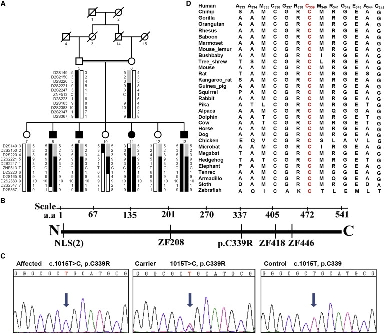
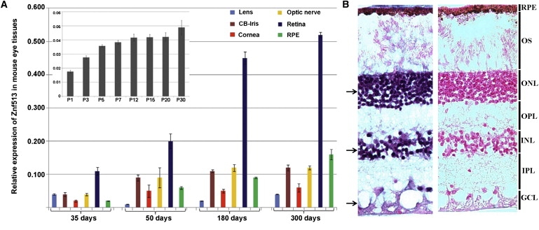
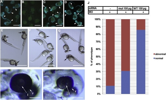
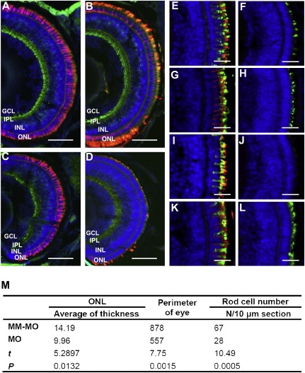
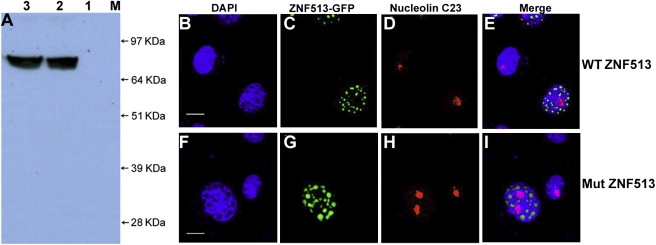
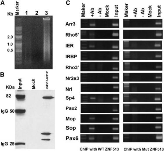
Retinitis pigmentosa (RP) is a phenotypically and genetically heterogeneous group of inherited retinal degenerations characterized clinically by night blindness, progressive constriction of the visual fields, and loss of vision, and pathologically by progressive loss of rod and then cone photoreceptors. Autosomal-recessive RP (arRP) in a consanguineous Pakistani family previously linked to chromosome 2p22.3-p24.1 is shown to result from a homozygous missense mutation (c.1015T>C [p.C339R]) in ZNF513, encoding a presumptive transcription factor. znf513 is expressed in the retina, especially in the outer nuclear layer, inner nuclear layer, and photoreceptors. Knockdown of znf513 in zebrafish reduces eye size, retinal thickness, and expression of rod and cone opsins and causes specific loss of photoreceptors. These effects are rescued by coinjection with wild-type (WT) but not p.C339R-znf513 mRNA. Both normal and p.C339R mutant ZNF513 proteins expressed in COS-7 cells localize to the nucleus. ChIP analysis shows that only the wild-type but not the mutant ZNF513 binds to the Pax6, Sp4, Arr3, Irbp, and photoreceptor opsin promoters. These results suggest that the ZNF513 p.C339R mutation is responsible for RP in this family and that ZNF513 plays a key role in the regulation of photoreceptor-specific genes in retinal development and photoreceptor maintenance.
2010 The American Society of Human Genetics. Published by Elsevier Inc. All rights reserved.
Figures
Similar articles
-
Whole-exome sequencing reveals ZNF408 as a new gene associated with autosomal recessive retinitis pigmentosa with vitreal alterations.Hum Mol Genet. 2015 Jul 15;24(14):4037-48. doi: 10.1093/hmg/ddv140. Epub 2015 Apr 16. Hum Mol Genet. 2015. PMID: 25882705
-
The manner of decay of genetically defective EYS gene transcripts in photoreceptor-directed fibroblasts derived from retinitis pigmentosa patients depends on the type of mutation.Stem Cell Res Ther. 2018 Oct 25;9(1):279. doi: 10.1186/s13287-018-1016-9. Stem Cell Res Ther. 2018. PMID: 30359287 Free PMC article.
-
Nonsense mutations in FAM161A cause RP28-associated recessive retinitis pigmentosa.Am J Hum Genet. 2010 Sep 10;87(3):376-81. doi: 10.1016/j.ajhg.2010.07.018. Epub 2010 Aug 12. Am J Hum Genet. 2010. PMID: 20705278 Free PMC article.
-
Non-syndromic retinitis pigmentosa.Prog Retin Eye Res. 2018 Sep;66:157-186. doi: 10.1016/j.preteyeres.2018.03.005. Epub 2018 Mar 27. Prog Retin Eye Res. 2018. PMID: 29597005 Review.
-
Variant haploinsufficiency and phenotypic non-penetrance in PRPF31-associated retinitis pigmentosa.Clin Genet. 2016 Aug;90(2):118-26. doi: 10.1111/cge.12758. Epub 2016 Mar 4. Clin Genet. 2016. PMID: 26853529 Review.
Cited by
-
Functional Genomics of the Retina to Elucidate its Construction and Deconstruction.Int J Mol Sci. 2019 Oct 4;20(19):4922. doi: 10.3390/ijms20194922. Int J Mol Sci. 2019. PMID: 31590277 Free PMC article. Review.
-
Deciphering the genetic architecture and ethnographic distribution of IRD in three ethnic populations by whole genome sequence analysis.PLoS Genet. 2021 Oct 18;17(10):e1009848. doi: 10.1371/journal.pgen.1009848. eCollection 2021 Oct. PLoS Genet. 2021. PMID: 34662339 Free PMC article.
-
Genetic characterization and disease mechanism of retinitis pigmentosa; current scenario.3 Biotech. 2017 Aug;7(4):251. doi: 10.1007/s13205-017-0878-3. Epub 2017 Jul 18. 3 Biotech. 2017. PMID: 28721681 Free PMC article. Review.
-
The Role of Inflammation in Retinal Neurodegeneration and Degenerative Diseases.Int J Mol Sci. 2021 Dec 30;23(1):386. doi: 10.3390/ijms23010386. Int J Mol Sci. 2021. PMID: 35008812 Free PMC article. Review.
-
Pathogenic mutations in TULP1 responsible for retinitis pigmentosa identified in consanguineous familial cases.Mol Vis. 2016 Jul 16;22:797-815. eCollection 2016. Mol Vis. 2016. PMID: 27440997 Free PMC article.
References
-
- Bird A.C. Retinal photoreceptor dystrophies LI. Edward Jackson Memorial Lecture. Am. J. Ophthalmol. 1995;119:543–562. - PubMed
-
- Bhatti M.T. Retinitis pigmentosa, pigmentary retinopathies, and neurologic diseases. Curr. Neurol. Neurosci. Rep. 2006;6:403–413. - PubMed
-
- Bunker C.H., Berson E.L., Bromley W.C., Hayes R.P., Roderick T.H. Prevalence of retinitis pigmentosa in Maine. Am. J. Ophthalmol. 1984;97:357–365. - PubMed
-
- Rivolta C., Sharon D., DeAngelis M.M., Dryja T.P. Retinitis pigmentosa and allied diseases: numerous diseases, genes, and inheritance patterns. Hum. Mol. Genet. 2002;11:1219–1227. - PubMed
-
- Grondahl J. Estimation of prognosis and prevalence of retinitis-pigmentosa and Usher Syndrome in Norway. Clin. Genet. 1987;31:255–264. - PubMed
Publication types
MeSH terms
Substances
Grants and funding
LinkOut - more resources
Full Text Sources
Molecular Biology Databases
