. 2010 Sep 10;87(3):371-5.
doi: 10.1016/j.ajhg.2010.08.001.

A focal epilepsy and intellectual disability syndrome is due to a mutation in TBC1D24

Affiliations

A focal epilepsy and intellectual disability syndrome is due to a mutation in TBC1D24

Mark A Corbett et al. Am J Hum Genet. .

Abstract

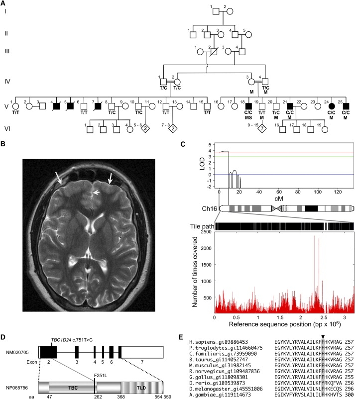
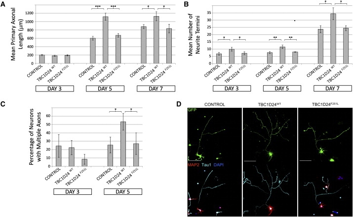
We characterized an autosomal-recessive syndrome of focal epilepsy, dysarthria, and mild to moderate intellectual disability in a consanguineous Arab-Israeli family associated with subtle cortical thickening. We used multipoint linkage analysis to map the causative mutation to a 3.2 Mb interval within 16p13.3 with a LOD score of 3.86. The linked interval contained 160 genes, many of which were considered to be plausible candidates to harbor the disease-causing mutation. To interrogate the interval in an efficient and unbiased manner, we used targeted sequence enrichment and massively parallel sequencing. By prioritizing unique variants that affected protein translation, a pathogenic mutation was identified in TBC1D24 (p.F251L), a gene of unknown function. It is a member of a large gene family encoding TBC domain proteins with predicted function as Rab GTPase activators. We show that TBC1D24 is expressed early in mouse brain and that TBC1D24 protein is a potent modulator of primary axonal arborization and specification in neuronal cells, consistent with the phenotypic abnormality described.


Figures

Figure 1
Figure 1
Mapping and Identification of a Mutation in TBC1D24 (A) Pedigree showing evidence of likely autosomal-recessive inheritance and consanguinity. Individuals used in homozygosity mapping are marked with M, the individual sequenced by next-generation sequencing is marked with S, and C/C, C/T, and T/T indicate TBC1D24 c.751 alleles. Solid squares indicate affected individuals. (B) Axial MRI image of V-25 that we interpreted as showing thickened cortex in the frontal poles with loss of gray-white matter definition, consistent with a developmental malformation (arrows). (C) Work flow for the identification of the mutation in TBC1D24: linkage homozygous-by-descent analysis (top) identified a single peak on chromosome 16p13.3 covering 9cM (or 3.2 Mb); the entire sequence, excluding repeats, was tiled as indicated by the black sections on the tile path; sequence coverage (bottom), determined by the number of times a base was covered by a sequence read, shows variation over the 3.2 Mb interval. (D) A schematic representation of the exonic structure of TBC1D24 and the domain structure of the TBC1D24 protein. The position of the c.751C>T, p.F251L mutation within the TBC domain (translated from exon 2) is shown. Exons are indicated as boxes, with the translated region shown in black. The TBC and TLD domains are shown. (E) A portion of a CLUSTALW multiple protein alignment of TBC1D24 orthologs shows that p.F251 (highlighted with gray background) is conserved in mammals, chicken, fruit fly, mosquito, and a highly similar tyrosine (Y) substitution in zebra fish. Orthologs of TBC1D24 were identified by tblastn search, and the Homologene database and alignments were performed with the EBI CLUSTALW server.
Figure 2
Figure 2
TBC1D24 Is a Potent Modulator of Neuronal Cell Morphology and Primary Axon Specification For all experiments: ED18.5 hippocampal neurons were isolated via established methods and then transfected with the Amaxa Mouse Neuron Nucleofector kit (Lonza). Cells were transfected with 1 μg of the supplied pmaxGFP vector alone (control) or in addition to 2 μg of either a commercially available expression vector RC210346 (NM_020705; OriGene Technologies) for the human TBC1D24 ORF (TBC1D24WT) or the same RC210346 vector with the c.751T>C mutation introduced by site-directed mutagenesis (TBC1D24F251L). Following transfection, neurons were plated onto poly-D-lysine (Sigma)-coated coverslips at a density of 1.25 × 10⁵ cells per well in Neurobasal media containing 2% (vol/vol) B27 (Invitrogen) and 10% fetal calf serum (FCS, Thermoscientific). Once cells were attached (∼4 hr), the FCS was removed and the cells were cultured for a further 3, 5, or 7 days, with half of the media changed every 4 days. Cells were fixed with 4% paraformaldehyde for 15 min at room temperature and then permeabilized, and nonspecific antigens were blocked with a solution of phosphate-buffered saline containing 1% Tween 20 (PBST) and 10% normal horse serum (Sigma). Primary antibodies were incubated overnight at 4°C at the following dilutions: chicken anti-MAP2 (1:2000, Chemicon, Millipore Bioscience Research Products) to identify dendrites or mouse anti-Tau1 (1:2000, Chemicon) to identify axons. Either donkey anti-mouse-Alexa647 (Invitrogen) or donkey anti-chicken-Cy3 (Chemicon) secondary antibodies were used at a dilution of 1:800. Nuclei were stained with DAPI as per the manufacturer's protocol (Invitrogen). Coverslips were mounted with Slowfade antifade mounting media (Invitrogen). p < 0.05, ∗∗p < 0.01, ∗∗∗p < 0.005 by Student's two-tailed t test, assuming equal variances. (A) Overexpression of TBC1D24WT but not TBC1D24F251L promotes axonal growth in cultured hippocampal neurons. Graph depicts average length of primary axons ± standard deviation (SD) from triplicate experiments measured with the “measure cumulative distances” plugin for the ImageJ software package (National Institutes of Health), with at least 20 measurements made for each treatment. (B) Overexpression of TBC1D24WT but not TBC1D24F251L promotes arborization of cultured hippocampal neurons. Graph depicts the average number of neurite termini per cell ± SD from triplicate experiments, with at least 10 neurons counted for each treatment. (C) Overexpression of TBC1D24WT but not TBC1D24F251L promotes ectopic axonal specification in hippocampal neurons after 5 days' culture. Graph shows average percentage of transfected cells with multiple axons ± SD from triplicate experiments, with at least 10 neurons counted for each treatment. (D) Example images of transfected neurons at 5 days of differentiation, showing either native GFP fluorescence or indirect immunofluorescent staining with Tau1, MAP2, and DAPI, as described. Scale bars represent 100 μM.

References

    1. Airaksinen E.M., Matilainen R., Mononen T., Mustonen K., Partanen J., Jokela V., Halonen P. A population-based study on epilepsy in mentally retarded children. Epilepsia. 2000;41:1214–1220. - PubMed
    1. Helbig I., Scheffer I.E., Mulley J.C., Berkovic S.F. Navigating the channels and beyond: Unravelling the genetics of the epilepsies. Lancet Neurol. 2008;7:231–245. - PubMed
    1. Mefford H.C., Eichler E.E. Duplication hotspots, rare genomic disorders, and common disease. Curr. Opin. Genet. Dev. 2009;19:196–204. - PMC - PubMed
    1. Tarpey P.S., Smith R., Pleasance E., Whibley A., Edkins S., Hardy C., O'Meara S., Latimer C., Dicks E., Menzies A. A systematic, large-scale resequencing screen of X-chromosome coding exons in mental retardation. Nat. Genet. 2009;41:535–543. - PMC - PubMed
    1. Albert T.J., Molla M.N., Muzny D.M., Nazareth L., Wheeler D., Song X., Richmond T.A., Middle C.M., Rodesch M.J., Packard C.J. Direct selection of human genomic loci by microarray hybridization. Nat. Methods. 2007;4:903–905. - PubMed

Publication types

MeSH terms