. 2010 Sep 3;142(5):714-25.
doi: 10.1016/j.cell.2010.07.038.

A C-type lectin collaborates with a CD45 phosphatase homolog to facilitate West Nile virus infection of mosquitoes

Affiliations

A C-type lectin collaborates with a CD45 phosphatase homolog to facilitate West Nile virus infection of mosquitoes

Gong Cheng et al. Cell. .

Abstract

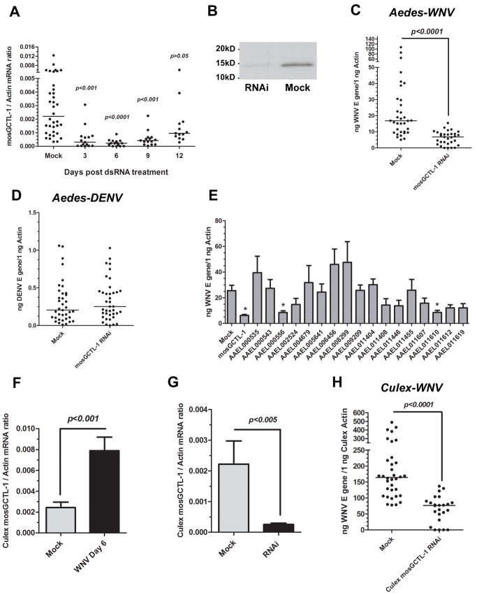
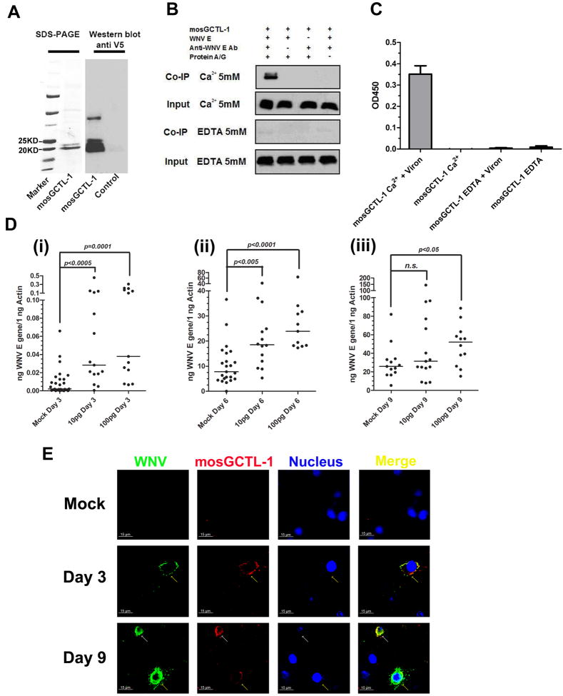
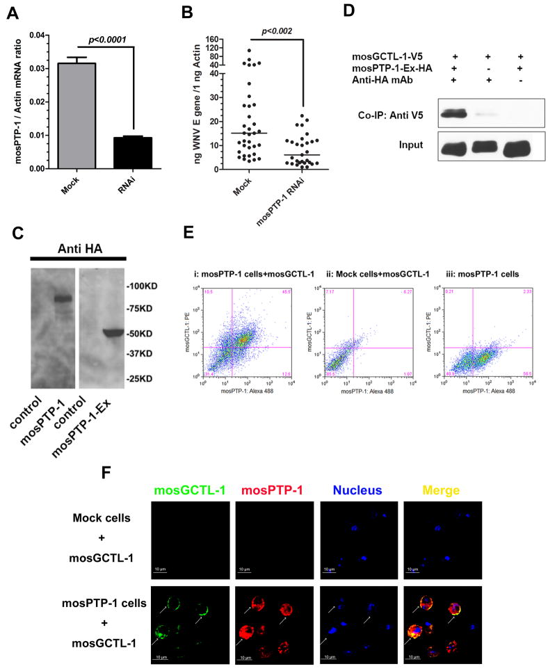
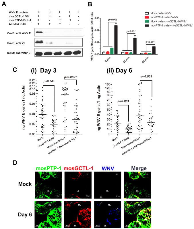
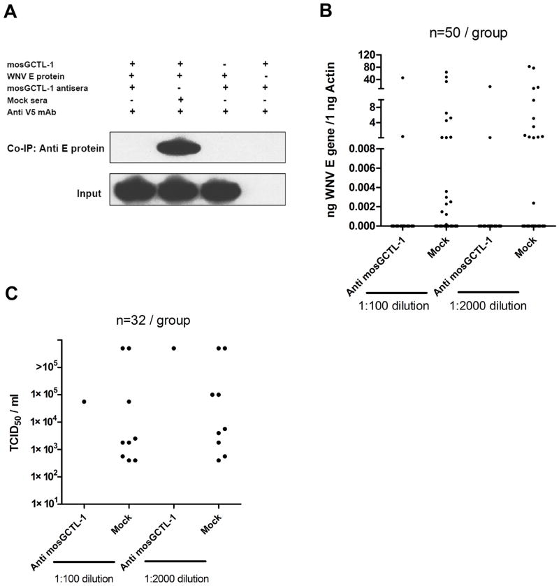
West Nile virus (WNV) is the most common arthropod-borne flavivirus in the United States; however, the vector ligand(s) that participate in infection are not known. We now show that an Aedes aegypti C-type lectin, mosGCTL-1, is induced by WNV, interacts with WNV in a calcium-dependent manner, and facilitates infection in vivo and in vitro. A mosquito homolog of human CD45 in A. aegypti, designated mosPTP-1, recruits mosGCTL-1 to enable viral attachment to cells and to enhance viral entry. In vivo experiments show that mosGCTL-1 and mosPTP-1 function as part of the same pathway and are critical for WNV infection of mosquitoes. A similar phenomenon was also observed in Culex quinquefasciatus, a natural vector of WNV, further demonstrating that these genes participate in WNV infection. During the mosquito blood-feeding process, WNV infection was blocked in vivo with mosGCTL-1 antibodies. A molecular understanding of flaviviral-arthropod interactions may lead to strategies to control viral dissemination in nature.


Figures

Figure 1
Figure 1. Effect of WNV infection on mosGCTL-1 expression
(A) Viral distribution in A. aegypti. WNV (1 × 10³ M.I.D50) was inoculated into the mosquito thorax. (B-E) mosGCTL-1 mRNA was induced by viral infection in whole A. aegypti (B), salivary glands (C), hemolymph (D), and midgut (E). Total RNA was isolated from various tissues or whole mosquitoes at 5 time points following viral infection. (A-E) The viral load and mosGCTL-1 mRNA levels were determined by Taqman® RT-QPCR, and normalized using A. aegypti actin (AAEL011197). WNV (1×10³ M.I.D50) was microinjected into each mosquito. Data are shown as the mean ± standard error (SEM). The experiments were repeated 3 times. (F) Immunoblot to detect mosGCTL-1 in WNV-infected mosquitoes. Three WNV infected or control mosquitoes were pooled and homogenized. The supernatant was then isolated, separated by SDS-PAGE, and probed with rabbit mosGCTL-1 antisera. UI, uninfected mosquitoes. I, WNV infected mosquitoes. dpi, days post infection. 50 μg of protein from mosquito lysates was loaded into each lane. Also see Figure S1 and Table S1.
Figure 2
Figure 2. The function of mosGCTL-1 in WNV infection
(A-B) mosGCTL-1 silencing. The mock group was treated with the same amount of GFP dsRNA. mosGCTL-1- or GFP-dsRNA-treated mosquitoes were used to isolate total RNA at several time points post dsRNA injection. mRNA levels were determined by SYBR Green® RT-QPCR (A). mosGCTL-1-dsRNA-treated or mock-treated mosquitoes were collected at 6 days post gene silencing. The supernatant separated by SDS-PAGE and probed with rabbit mosGCTL-1 antisera (B). (C-D) Silencing mosGCTL-1 impairs WNV (C), but not dengue virus (D), infection. The viral burden was examined at day 6 post-infection. 10 M.I.D50 WNV or dengue virus was used to challenge mosquitoes. The viral load was determined by Taqman® RT-QPCR, and normalized using A. aegypti actin. The result shown was representative of 4 independent experiments. (E) The role of the mosGCTL paralogues in WNV infection. The sample number was no less than 12 mosquitoes in each group. The viral burden was determined by Taqman® RT-QPCR, and normalized with A. aegypti actin. *, p<0.05. The results were pooled from 2 independent experiments. (F) WNV induces mosGCTL-1 homologue expression in C. quinquefasciatus. WNV-infected and mock mosquitoes were collected at 6 days post infection. Culex mosGCTL-1 mRNA was determined by SYBR Green® RT-QPCR, and normalized using C. quinquefasciatus actin (CPIJ012570). The experiment was repeated 3 times with similar results. (G) Efficiency of Culex mosGCTL-1 RNA interference. The mock group was treated with the same amount of GFP dsRNA. mRNA levels were determined by SYBR Green® RT-QPCR. (H) Silencing Culex mosGCTL-1 decreases the WNV burden in C. quinquefasciatus. The viral burden was determined by Taqman® RT-QPCR. The results were combined from 3 independent experiments. (A, C-H) Statistical analysis was done with the Mann-Whitney test in all experiments. Each dot represents the mRNA levels in an individual mosquito. The horizontal line depicts the medians. (E-G) Data are shown as the mean ± standard error (SEM). Also see Figure S2 and Table S2.
Figure 3
Figure 3. mosGCTL-1 interacts with WNV
(A) Recombinant mosGCTL-1, produced in Drosophila cells, and purified using a Ni-His column (Left panel). Glycosylated mosGCTL-1 was detected with a V5-HRP mAb (Right panel). The control was the supernatant of mock-infected S2 cells. (B) mosGCTL-1 interacted with WNV E protein in a co-immunoprecipitation assay. The protein complex was pulled down with a flavivirus E mAb and probed with anti-V5-HRP mAb. The experiments were repeated 3 times. (C) mosGCTL-1 captured West Nile virions by ELISA. The interaction was determined using a flavivirus E mAb. Data are expressed as the mean ± standard error from 3 independent experiments. (D) Co-microinjecting the purified mosGCTL-1 with WNV enhanced virus infection in A. aegypti. The various amount of purified mosGCTL-1 were pre-mixed with WNV (10 M.I.D50 per mosquito) for 1 hour at 4°C. The mixture was microinjected into mosquito and compared to the control group inoculated with the same amount WNV. The mosquitoes were collected at 3 days (i), 6 days (ii) and 9 days (iii) post WNV inoculation. Total RNA was isolated to determine the viral burden by Taqman® RT-QPCR, normalized using A. aegypti actin. Each dot represents the mRNA level in one mosquito. The horizontal line represents the medians. n.s., nonsignificance (p > 0.05). The Mann-Whitney test was used for analysis. Three independent experiments yielded similar data. (E) The association between mosGCTL-1 and WNV in A.aegypti hemolymph. Hemolymph was collected from WNV-infected or mock-infected mosquitoes for immunofluorescence staining. WNV E protein was stained with anti-mouse IgG Alexa-488 (Green), and mosGCTL-1 was identified using anti-rabbit IgG Alexa-546 (Red). Nuclei were stained blue with To-Pro-3 iodide. The white arrow represents the induced expression of mosGCTL-1 in WNV-infected hemocytes. The yellow arrows show the infected hemocytes. Images were examined using a Zeiss LSM 510 meta confocal 63×objective lens. Also see Figure S3.
Figure 4
Figure 4. mosPTP-1 captures mosGCTL-1 onto the cell surface
(A) RNAi efficiency for the mosPTP-1 gene at 6 days post dsRNA treatment. The amount of mosPTP-1 mRNA was determined by SYBR Green® RT-QPCR, and normalized using A. aegypti actin. Data are represented as the mean ± standard error. (B) Silencing mosPTP-1 decreased WNV infection. The viral burden was measured at day 6. 10 M.I.D50 of WNV was injected into each mosquito. The viral load was determined by Taqman® RT-QPCR, and normalized using A. aegypti actin. Statistical analysis was done with the Mann-Whitney test. The horizontal line depicts the medians. The result shown is a combination of 3 independent experiments. (C) Expression of recombinant mosPTP-1 and mosPTP-1-Ex in S2 cells. mosPTP-1 and mosPTP-1-Ex genes were isolated from cDNA library of A. aegypti, and expressed as recombinant proteins with an HA tag at the N-terminus. The left panel is mosPTP-1 and the right panel is mosPTP-1-EX, probed by anti-HA tag mAb in western-blot. The control was the products from S2 cells transfected with mock DNA vector. (D) mosGCTL-1 interacts with mosPTP-1-Ex peptide by co-immunoprecipitation. The protein complex was pulled down with a rabbit HA antibody, and probed with a V5-HRP mAb. The experiments were repeated 3 times. (E) mosPTP-1 captured mosGCTL-1 to the cell surface by flow cytometry. A stable cell line was generated to express mosPTP-1 in S2 cells. The purified mosGCTL-1 was inoculated with mosPTP-1 expressing cells at 4°C. An empty DNA vector transfected stable S2 cell line was used as the control cells. The interaction between mosPTP-1 and mosGCTL-1 was investigated by FACS. mosPTP-1 was stained by Alexa-488; mosGCTL-1 was stained by Phycoerythrin (PE). Three independent experiments yielded similar results, and one representative study is shown in this figure. (F) Confocal microscopy to examine for mosPTP-1 and mosGCTL-1. mosGCTL-1 was stained with Alexa-488 (Green) and mosPTP-1 was identified with Alexa-546 (Red). Nuclei were stained by To-Pro-3 iodide (blue). The images were collected using a Zeiss LSM 510 meta confocal microscopy 63×objective lens. The arrows represent the overlap between mosPTP-1 and mosGCTL-1. Also see Figure S4 and Table S3.
Figure 5
Figure 5. The mosGCTL-1/mosPTP-1 pathway in WNV infection
(A) mosGCTL-1 associated with WNV E protein bound to mosPTP-1-Ex. The protein complex was pulled down with an HA antibody and probed with V5-HRP and flavivirus E protein antibody. The experiment was repeated 3 times. (B) mosPTP-1-expressing cells recruit WN virions in the presence of mosGCTL-1. mosGCTL-1 and WNV were added to cells and incubated at 4°C for 1 hr, for membrane attachment. Cells were gently washed 3 times using cold PBS, and then moved to room temperature. At different time points, 0, 15 and 60 min, cells were collected for total RNA isolation. Control cells were the empty DNA vector transfected stable S2 cell line. Viral attachment was determined by SYBR Green® RT-QPCR, and normalized with Drosophila actin 5C (CG4027). Statistical analysis was done with ANOVA. Data are represented as the mean ± standard error. The results are representative of 3 independent experiments. (C) Silencing mosPTP-1 impairs the function of mosGCTL-1. The mosPTP-1 gene was knocked down using dsRNA treatment. Then the mosGCTL-1/WNV mixture was inoculated at 3 days post RNAi-silencing. The virus burden was measured at 3 (i) and 6 (ii) days after the introduction of virus by Taqman® RT-QPCR. 10 M.I.D50 WNV and 100 pg mosGCTL-1 were inoculated into each mosquito. Each dot represents the mRNA level in an individual mosquito. Statistical analysis was done using the Mann-Whitney test. The horizontal line depicts the medians. The result shown is the combination of 5 independent experiments. (D) Immunostaining of mosGCTL-1, mosPTP-1 and WNV in A. aegypti salivary glands. mosPTP-1 was stained with anti-mouse IgG Alexa-488 (Green); mosGCTL-1 was identified by anti-rabbit IgG Alexa-546 (Red); WNV E protein was probed with horse anti-E protein IgG and detected by anti-horse IgG Alexa-633 (Blue). The arrows show the sites of overlap between mosGCTL-1, mosPTP-1 and WNV at 6 days post infection. LL, Lateral Lobe; ML, Median Lobe in female A.aegypti salivary glands. Images were examined using a Zeiss LSM 510 meta confocal 25×objective lens. Also see Figure S5.
Figure 6
Figure 6. mosGCTL-1 antiserum interferes with WNV infection of mosquitoes
(A) mosGCTL-1 antisera blocked the interaction between mosGCTL-1 and WNV E protein. mosGCTL-1 antisera or mock sera was diluted 1000-fold. The protein complex was pulled down with a V5 mAb and WNV E protein was detected using an E protein antibody. (B-C) mosGCTL-1 antisera interrupted WNV infection during the blood meal. The antisera or control sera were diluted 100- or 2000-fold with fresh whole blood containing 5×10⁶ pfu/ml WNV. Membrane blood-feeding was then performed using a Hemotek®. Seven days later, mosquitoes were sacrificed to determine the infectivity rate by Taqman® RT-QPCR (B) and TCID50 (C). Each group included 50 mosquitoes in the QPCR assay, and 32 mosquitoes in TCID50 assay. One dot corresponds to a mosquito. n, the number of mosquitoes in each group. The result is representative of 3 independent experiments.

Similar articles

Cited by

References

    1. Alonso A, et al. Protein tyrosine phosphatases in the human genome. Cell. 2004;117:699–711. - PubMed
    1. Arnold JN, Dwek RA, Rudd PM, Sim RB. Mannan binding lectin and its interaction with immunoglobulins in health and in disease. Immunol Lett. 2006;106:103–110. - PubMed
    1. Bai FW, et al. Antiviral peptides targeting the West Nile virus envelope protein. J Virol. 2007;81:2047–2055. - PMC - PubMed
    1. Baldwin TA, Ostergaard HL. Developmentally regulated changes in glucosidase II association with, and carbohydrate content of, the protein tyrosine phosphatase CD45. J Immunol. 2001;167:3829–3835. - PubMed
    1. Blandin S, Shiao SH, Moita LF, Janse CJ, Waters AP, Kafatos FC, Levashina EA. Complement-like protein TEP1 is a determinant of vectorial capacity in the malaria vector Anopheles gambiae. Cell. 2004;116:661–670. - PubMed

Publication types