. 2010 Sep 24;39(6):925-38.
doi: 10.1016/j.molcel.2010.08.011.

The nuclear-retained noncoding RNA MALAT1 regulates alternative splicing by modulating SR splicing factor phosphorylation

Affiliations

The nuclear-retained noncoding RNA MALAT1 regulates alternative splicing by modulating SR splicing factor phosphorylation

Vidisha Tripathi et al. Mol Cell. .

Abstract

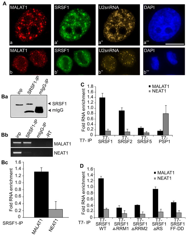
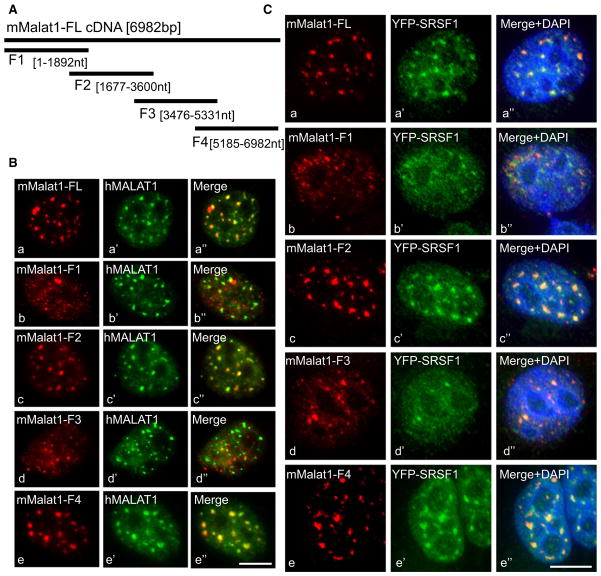
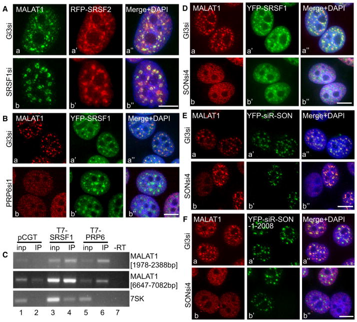
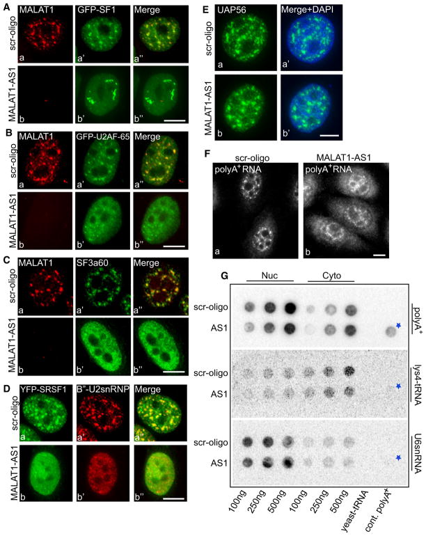
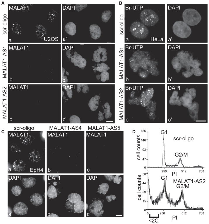
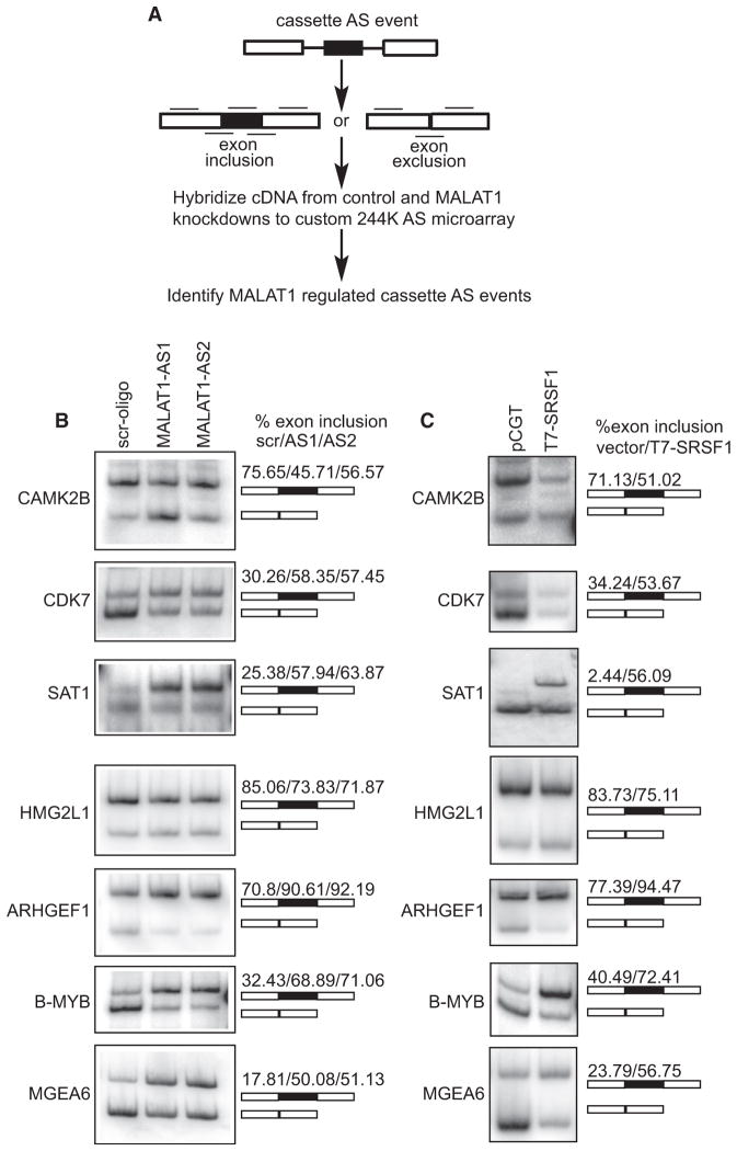
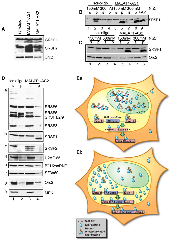
Alternative splicing (AS) of pre-mRNA is utilized by higher eukaryotes to achieve increased transcriptome and proteomic complexity. The serine/arginine (SR) splicing factors regulate tissue- or cell-type-specific AS in a concentration- and phosphorylation-dependent manner. However, the mechanisms that modulate the cellular levels of active SR proteins remain to be elucidated. In the present study, we provide evidence for a role for the long nuclear-retained regulatory RNA (nrRNA), MALAT1 in AS regulation. MALAT1 interacts with SR proteins and influences the distribution of these and other splicing factors in nuclear speckle domains. Depletion of MALAT1 or overexpression of an SR protein changes the AS of a similar set of endogenous pre-mRNAs. Furthermore, MALAT1 regulates cellular levels of phosphorylated forms of SR proteins. Taken together, our results suggest that MALAT1 regulates AS by modulating the levels of active SR proteins. Our results further highlight the role for an nrRNA in the regulation of gene expression.


Figures

Figure 1
Figure 1. MALAT1 Localizes to Nuclear Speckles and Interacts with SR Proteins
(A) Co-RNA-FISH using probes against MALAT1 (Aa and Ab), U2 snRNA (Aa″ and Ab″) and immunostaining using an antibody against SRSF1 (Aa′ and Ab′) in interphase (Aa–Aa‴) and mitotic telophase HeLa cells (Ab–Ab‴). The DNA is counterstained with DAPI (blue; Aa‴ and Ab‴). The bar represents 10 μm. (B) (Ba) Immunoprecipitation from HeLa cells using SRSF1 antibody or mouse serum followed by immunoblot with SRSF1 antibody. (Bb and Bc) RT-PCR or qPCR from the IP samples using MALAT1- or NEAT1-specific primers. (C) qPCR from T7-IP (T7-SRSF1-WT-, T7-SRSF2-, T7-SRSF5-, and T7-PSP1-expressing cells) using MALAT1- or NEAT1-specific primers. (D) qPCR from T7-IP (T7-SRSF1-WT, T7-SRSF1ΔRRM1, ΔRRM2, ΔRS, FF-DD-expressing cells) using MALAT1- or NEAT1-specific primers. Error bars in (Bc), (C), and (D) represent mean ± SD of three independent experiments. See also Figure S1.
Figure 2
Figure 2. MALAT1 RNA Contains Two Independent Nuclear Speckle-Localizing Motifs
(A) Schematic representation of full-length mouse MALAT1 (6982 bp) and mutant constructs (F1–F4). (B) Co-RNA FISH using mouse (Ba–Be)- and human (Ba′–Be′)-specific MALAT1 probes in HeLa cells that were transiently transfected with full-length (a) and mutant mMALAT1 constructs (F1 [Bb], F2 [Bc], F3 [Bd], and F4 [e]). (C) RNA FISH using mMALAT1 probe (Ca–Ce) in HeLa cells expressing YFP-SRSF1 (Ca′–Ce′) that were transiently transfected with full-length mMALAT1 cDNA (Ca) and mutant constructs (F1 [b], F2 [c], F3 [d], and F4 [e]). The bar represents 10 μm. See also Figure S2.
Figure 3
Figure 3. Nuclear Speckle-Associated Splicing Factors PRP6 and SON Influence the Distribution of MALAT1 in Nuclear Speckles
(A) MALAT1 RNA-FISH (Aa and Ab) in control luciferase siRNA (GL3si; a–a″) and SRSF1 siRNA (b–b″)-treated HeLa cells expressing RFP-SRSF2 (a′–b′). (B) MALAT1 RNA-FISH in control (Ba–Ba″) and PRP6 siRNA-1 (Bb–Bb″)-treated YFP-SRSF1 (Ba′–Bb′) stably expressing HeLa cells. (C) RT-PCR from T7-antibody IP samples (lanes 2, 4, and 6) of HeLa cells expressing T7 (pCGT vector) alone (lanes 1 and 2), T7-SRSF1 (lanes 3 and 4) and T7-PRP6 (lanes 5 and 6), using MALAT1 primers. RT-PCR using 7SK primers were performed as a negative control. (D) MALAT1 RNA-FISH (Da and Db) in control (Da–Da″) and SON siRNA4 (Db–Db″)-treated YFP-SRSF1 (Da′ and Db′)-expressing HeLa cells. (E) MALAT1 RNA-FISH in control (Ea–Ea″) and SON siRNA4 (Eb–Eb″)-treated HeLa cells, which were transiently expressing siRNA4 refractory YFP-SON-FL (YFP-siR-SON; Ea′ and Eb′). Note that exogenously expressed SON restored the speckle distribution of MALAT1 (Eb–Eb″). (F) MALAT1 RNA-FISH in control (Fa–Fa″) and SON siRNA4 (Fb–Fb″)-treated HeLa cells, which were transiently expressing siRNA4 refractory YFP-SON mutant (green; YFP-siR-SON-1-2008; Fa′ and Fb′). The bars in all the figures represent 10 μm. See also Figure S3.
Figure 4
Figure 4. MALAT1 Is Not Required for Nuclear Speckle Structural Integrity but Influences the Distribution of Splicing Factors to Nuclear Speckles
(A and B) MALAT1 RNA-FISH in control (scr-oligo, a–a″) and MALAT1 antisense oligo-treated (48 hr post antisense treatment; b–b″) HeLa cells expressing GFP-SF1 (A) or GFP-U2AF65 (B). (C) MALAT1 RNA-FISH and SF3a60 immunolocalization in control (Ca–Ca″) and MALAT1-depleted (Cb–Cb″) HeLa cell. (D) YFP-SRSF1 and B″-U2snRNP immunolocalization in scr-oligo (Da–Da″) and MALAT1-depleted (Db–Db″) HeLa cell. (E) UAP56 immunolocalization in control (Ea–Ea′) and MALAT1-depleted (Eb–Eb′) HeLa cell. (F) RNA-FISH using oligo-dT probe in control (Fa) and MALAT1 antisense oligonucleotides-treated (Fb) HeLa cells. The bars in all the figures represent 10 μm. (G) RNA slot blot using oligo dT probe in control (scr-oligo) and MALAT1 antisense oligonucleotide-treated (AS1) HeLa cell nuclear (nuc) and cytoplasmic (cyto) extracts showed increased levels of cytoplasmic poly(A)+ RNA upon MALAT1 depletion (fold change, MALAT1 AS1/scr 100 ng = 0.63; 250 ng = 1.36; 500 ng = 1.6). See also Figure S4. The asterisk represents poly(A)+ RNA from control cells.
Figure 5
Figure 5. MALAT1 Depletion Results in the Fragmentation of Cell Nuclei
(A and C) MALAT1 RNA-FISH in U2OS (A) and EpH4 (C) cells that were transfected with control (scr-oligo; a–a′), human specific (A, AS1 and AS2; b–b′ and c–c′) or mouse-specific (C; AS4 and AS5; b–b′ and c–c′) MALAT1 antisense oligonucleotides (72 hr posttransfection). (B) In vivo Br-UTP transcription analyses in control (Ba–Ba′) and MALAT1 antisense oligo-transfected (Bb–Bb′ and Bc–Bc′) HeLa cells. The bars in (A), (B), and (C) represent 10 μm. (D) Flow cytometric analyses revealed increased cell death (<2C DNA content) with increased G2/M population in MALAT1-depleted HeLa cells. See also Figure S5.
Figure 6
Figure 6. MALAT1 Regulates AS of Pre-mRNAs
(A) Schematic representation of the method used for the global analysis of MALAT1-regulated AS. Black lines designate exon body and exon junction probes used for analyzing AS on a custom 244K Agilent microarray. (B and C) RT-PCR analysis using primers specific to exons in MALAT1 or to constitutive exons flanking SRSF1-regulated alternative exons in CAMK2B, CDK7, SAT1, HMG2L1, ARHGEF1, B-MYB, and MGEA6 mRNAs indicate changes in AS in MALAT1-depleted (6B) and SRSF1-overexpressed (6C) HeLa cells. Alternative exon-included (upper band) and -excluded bands (lower band) and the percentage change of alternative exon inclusion between scr. oligo/MALAT1-AS oligos (AS1 and AS2) observed is shown. See also Table S1.
Figure 7
Figure 7. MALAT1 Modulates the Cellular Levels and Phosphorylation of SR Proteins
(A) Immunoblot assays in control (scr) and MA-LAT1-depleted total cell extracts (AS1 and AS2) revealed increased levels of SRSF1 and SRSF2 in MALAT1-depleted cells. Relative fold increase in the levels of SR proteins in MALAT1-depleted cells: SRSF1, AS1 2.07 and AS2 3.13; SRSF2, AS1 3.15 and AS2 1.52. Note that the MALAT1-depleted cell extracts contained extra bands of SR proteins that show fast mobility. (B and C) Control (lanes 1–4) and MALAT1 anti-sense oligonucleotides (B, AS1; and C, AS2) transfected (lanes 5–8) HeLa cells were extracted using 150 and 300 mM NaCl salt into soluble supernatant (s; lanes 1, 3, 5, and 7) and insoluble chromatin pellet (p; lanes 2, 4, 6, and 8) fractions and immunoblotted using SRSF1 antibody. Lane 9 (B) represents total cell extract treated with Antarctic phosphatase (+AP). (D) Immunoblot analyses of control and MALAT1-depleted extracts (300 mM NaCl extracted) with 3C5 (Da), SRSF1 (Db), SRSF2 (Dc), U2AF-65 (Dd), B″-U2snRNP (De), SF3a60 (Df) antibodies. Orc2 and MEK were used as loading controls. (E) Hypothetical model depicting the role of MA-LAT1 in AS regulation. (Ea) In normal cells, MA-LAT1, by associating with SR proteins in nuclear speckles and in the nucleoplasm, regulates their recruitment to the pre-mRNA, thereby regulating AS. Here we have shown SAT1 pre-mRNA as an example that undergoes alternate exon exclusion in normal cells. However, in MALAT1-depleted cells (Eb), cellular levels of SR proteins are increased and are also present predominantly in the dephosphorylated form, resulting in changes in AS of pre-mRNA. In case of SAT1 pre-mRNA, MALAT1 depletion results in the inclusion of an alternative exon containing weak splice sites. See also Figure S6.

Comment in

Similar articles

Cited by

References

    1. Amaral PP, Dinger ME, Mercer TR, Mattick JS. The eukaryotic genome as an RNA machine. Science. 2008;319:1787–1789. - PubMed
    1. Blencowe BJ. Alternative splicing: new insights from global analyses. Cell. 2006;126:37–47. - PubMed
    1. Bourgeois CF, Lejeune F, Stevenin J. Broad specificity of SR (serine/arginine) proteins in the regulation of alternative splicing of pre-messenger RNA. Prog Nucleic Acid Res Mol Biol. 2004;78:37–88. - PubMed
    1. Caceres JF, Krainer AR. Functional analysis of pre-mRNA splicing factor SF2/ASF structural domains. EMBO J. 1993;12:4715–4726. - PMC - PubMed
    1. Caceres JF, Misteli T, Screaton GR, Spector DL, Krainer AR. Role of the modular domains of SR proteins in subnuclear localization and alternative splicing specificity. J Cell Biol. 1997;138:225–238. - PMC - PubMed

Publication types

MeSH terms