. 2010 Sep 7;107(36):15921-6.
doi: 10.1073/pnas.1010209107. Epub 2010 Aug 23.

Differentiated Parkinson patient-derived induced pluripotent stem cells grow in the adult rodent brain and reduce motor asymmetry in Parkinsonian rats

Affiliations

Differentiated Parkinson patient-derived induced pluripotent stem cells grow in the adult rodent brain and reduce motor asymmetry in Parkinsonian rats

Gunnar Hargus et al. Proc Natl Acad Sci U S A. .

Abstract

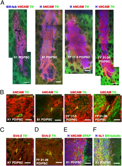
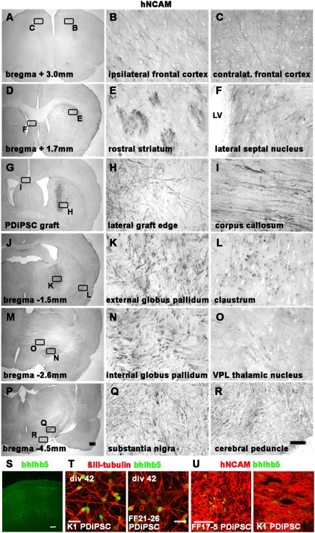
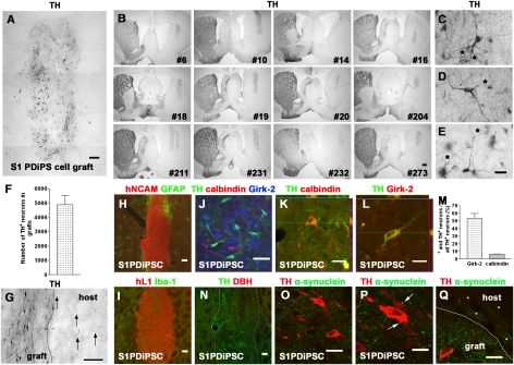
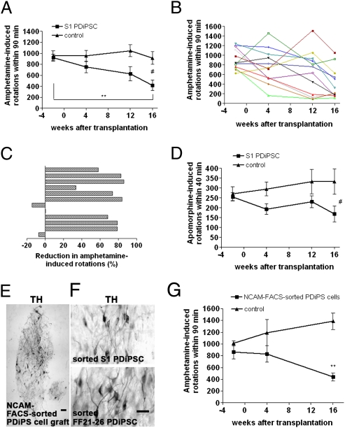
Recent advances in deriving induced pluripotent stem (iPS) cells from patients offer new possibilities for biomedical research and clinical applications, as these cells could be used for autologous transplantation. We differentiated iPS cells from patients with Parkinson's disease (PD) into dopaminergic (DA) neurons and show that these DA neurons can be transplanted without signs of neurodegeneration into the adult rodent striatum. The PD patient iPS (PDiPS) cell-derived DA neurons survived at high numbers, showed arborization, and mediated functional effects in an animal model of PD as determined by reduction of amphetamine- and apomorphine-induced rotational asymmetry, but only a few DA neurons projected into the host striatum at 16 wk after transplantation. We next applied FACS for the neural cell adhesion molecule NCAM on differentiated PDiPS cells before transplantation, which resulted in surviving DA neurons with functional effects on amphetamine-induced rotational asymmetry in a 6-OHDA animal model of PD. Morphologically, we found that PDiPS cell-derived non-DA neurons send axons along white matter tracts into specific close and remote gray matter target areas in the adult brain. Such findings establish the transplantation of human PDiPS cell-derived neurons as a long-term in vivo method to analyze potential disease-related changes in a physiological context. Our data also demonstrate proof of principle of survival and functional effects of PDiPS cell-derived DA neurons in an animal model of PD and encourage further development of differentiation protocols to enhance growth and function of implanted PDiPS cell-derived DA neurons in regard to potential therapeutic applications.


Conflict of interest statement

The authors declare no conflict of interest.

Figures

Fig. 1.
Fig. 1.
PD patient-derived DA neurons survive after transplantation into the adult striatum of unlesioned rats. (A) Assembled images of K1, S1, FF 17–5, and FF 21–26 PDiPS cell grafts immunostained for human NCAM (red), TH (green), βIII-tubulin (blue), or Hoechst (H; blue) 4 wk after intrastriatal transplantation of 200,000 cells. A high number of neurons was present in all grafts. (B) Z-stacks of three confocal images of 1 μm thickness show coexpression of TH (green) and hNCAM (red) in engrafted DA neurons derived from all four PDiPS cell lines 4 wk after transplantation. (C and D) Stacked confocal images of DA neurons coexpressing Girk-2 (red) and TH (green) 12 wk after transplantation. (E) Immunostaining for hNCAM (red), GFAP (green), and H (blue) showing moderate astrogliosis around grafts. (F) Immunostaining for human L1 (red), βIII-tubulin (green) and H (blue) showing a high number of neurons at the graft–host interface. (Scale bars: 100 μm in A, E, and F; 20 μm in BD.)
Fig. 2.
Fig. 2.
Engrafted PD patient–derived neurons send out fibers to close and remote target areas in the adult unlesioned rodent brain. (AR) Photomicrographs of hNCAM-stained brain sections 4 wk after engraftment of S1 PDiPS cells representing the axonal outgrowth pattern of engrafted PDiPS cell-, non-PDiPS cell–, and hES cell-derived neurons. Graft-derived axons project along white matter tracts (E, I, and R) to specific gray matter zone target areas in the adult rodent brain. The boxed areas (Left) are also shown in higher magnification (Right). (Scale bars: 25 μm, Right; 500 μm, Left.) LV, lateral ventricle; VPL, ventroposterolateral. (S) Immunostaining of the adult rat somatosensory cortex for the cortical marker bhlhb5. (Scale bar: 100 μm.) (T) Immunostainings of differentiated PDiPS cells at day 42 in vitro for βIII-tubulin (red) and bhlhb5 (green). (Scale bar: 20 μm.) (U) Immunostainings of differentiated PDiPS cells for hNCAM (red) and bhlhb5 (green) 4 wk after transplantation. (Scale bar: 50 μm.)
Fig. 3.
Fig. 3.
PDiPS cell-derived DA neurons survive at high numbers after transplantation into the striatum of 6-OHDA–lesioned rats. (AE and G) Photomicrographs of engrafted differentiated S1 PDiPS cells immunostained for TH 16 wk after transplantation of 400,000 cells into 6-OHDA–lesioned rats. (A) TH+ DA neurons were present at high numbers throughout the PDiPS cell grafts. Three images were assembled for graft reconstruction. (B) Low-power photomicrographs show that all 12 transplanted rats contained TH+ grafts in the DA-depleted striatum. Numbers indicate individual rats. (CE) DA neurons with fiber arborization and branching (asterisks) in grafts. (F) Quantification of DA neurons in PDiPS cell grafts. (G) Some graft-derived TH+ fibers (arrows) project into the host lesioned striatum. (H) Immunostainings of grafts for hNCAM (red) and GFAP (green) and (I) for hL1 (red) and Iba-1 (green) show low astroglial and microglial reaction around the grafts. (N) Immunostaining of engrafted cells for TH (green) and DBH (red) showing DA, although not noradrenergic, neurons in grafts. (JM) Girk-2 and calbindin were expressed in engrafted DA neurons. (J) Immunostaining of engrafted cells for TH (green), calbindin (red), and Girk-2 (blue). (K and L) Z-stacks of three confocal images show coexpression of (K) TH (green) and calbindin (red) or (L) TH (green) and Girk-2 (red) in engrafted DA neurons. (M) Quantification of stainings for TH, calbindin, and Girk-2 in PDiPS cell grafts. (OQ) Immunostainings for TH (red) and α-synuclein (green; human-specific antibody) show a punctate synaptic expression of α-synuclein on engrafted TH+ neurons (arrows, P) and in the host striatum (asterisks, Q). (Scale bars: 10 μm in P; 20 μm in K, L, O, and Q; 25 μm in CE; 50 μm in J; 100 μm in A, G, H, I, and N; 500 μm in B.)
Fig. 4.
Fig. 4.
Reduced motor asymmetry of 6-OHDA–lesioned rats after intrastriatal transplantation of differentiated PDiPS cells. (AC) Amphetamine-induced rotations of S1 PDiPS cell–transplanted rats (n = 12) significantly declined over time (**P < 0.01) and were significantly less compared with control rats (n = 9) 16 wk after transplantation (#P < 0.05). (B) Number of rotations of each rat over time. (C) The percentage of the reduction in rotations 16 wk after transplantation. (D) Apomorphine-induced rotations of rats over time reveal a significantly decreased number of rotations in PDiPS cell–treated rats 16 wk after transplantation compared with control rats (#P < 0.05). (EG) Differentiated S1 and FF21-26 PDiPS cells were FACS-sorted for NCAM and subsequently engrafted into five 6-OHDA–lesioned rats (S1, n = 2; FF21-26, n = 3). (E and F) Photomicrographs of TH+ DA neurons 16 wk after transplantation. Three images were assembled in panel E for graft reconstruction. (Scale bars: 50 μm in E; 25 μm in F.) (G) The number of amphetamine-induced rotations of rats engrafted with sorted PDiPS cells was significantly lower compared with control rats (n = 5) 16 wk after transplantation (**P < 0.01). Graphs show mean values ± SEM. Two-way ANOVA with post hoc Tukey test was performed for statistical analysis.

References

    1. Takahashi K, Yamanaka S. Induction of pluripotent stem cells from mouse embryonic and adult fibroblast cultures by defined factors. Cell. 2006;126:663–676. - PubMed
    1. Saha K, Jaenisch R. Technical challenges in using human induced pluripotent stem cells to model disease. Cell Stem Cell. 2009;5:584–595. - PMC - PubMed
    1. Wernig M, et al. Neurons derived from reprogrammed fibroblasts functionally integrate into the fetal brain and improve symptoms of rats with Parkinson's disease. Proc Natl Acad Sci USA. 2008;105:5856–5861. - PMC - PubMed
    1. Raya A, et al. Disease-corrected haematopoietic progenitors from Fanconi anaemia induced pluripotent stem cells. Nature. 2009;460:53–59. - PMC - PubMed
    1. Park IH, et al. Disease-specific induced pluripotent stem cells. Cell. 2008;134:877–886. - PMC - PubMed

Publication types