. 2010 Aug 31;107(35):15643-8.
doi: 10.1073/pnas.1003878107. Epub 2010 Aug 23.

Noninvasive method for assessing the human circadian clock using hair follicle cells

Affiliations

Noninvasive method for assessing the human circadian clock using hair follicle cells

Makoto Akashi et al. Proc Natl Acad Sci U S A. .

Abstract

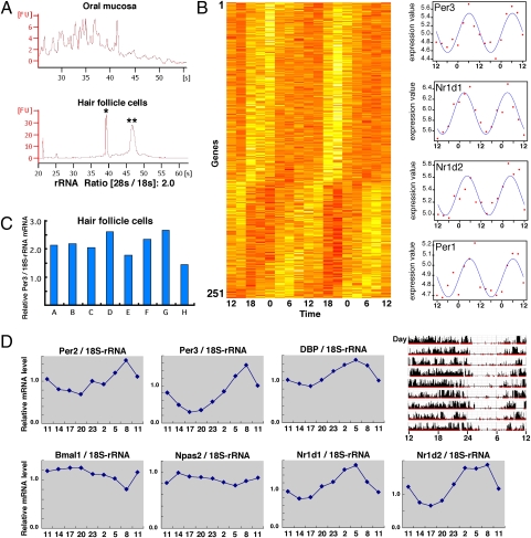
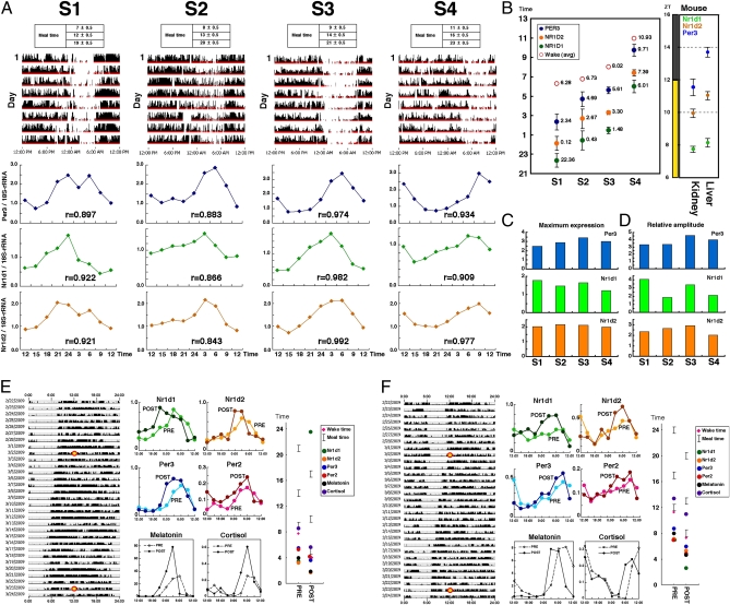
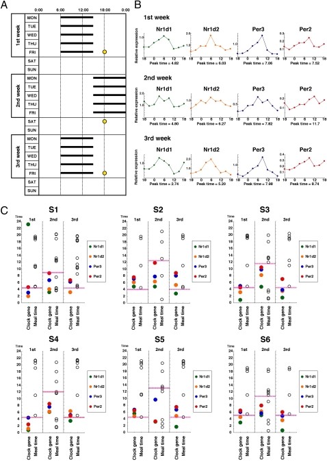
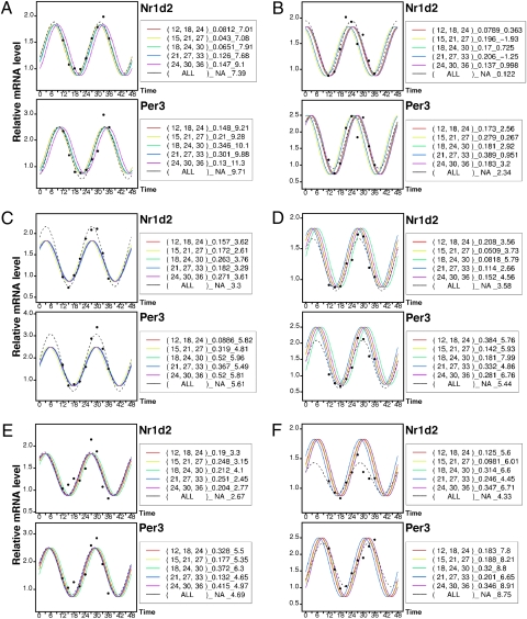
A thorough understanding of the circadian clock requires qualitative evaluation of circadian clock gene expression. Thus far, no simple and effective method for detecting human clock gene expression has become available. This limitation has greatly hampered our understanding of human circadian rhythm. Here we report a convenient, reliable, and less invasive method for detecting human clock gene expression using biopsy samples of hair follicle cells from the head or chin. We show that the circadian phase of clock gene expression in hair follicle cells accurately reflects that of individual behavioral rhythms, demonstrating that this strategy is appropriate for evaluating the human peripheral circadian clock. Furthermore, using this method, we indicate that rotating shift workers suffer from a serious time lag between circadian gene expression rhythms and lifestyle. Qualitative evaluation of clock gene expression in hair follicle cells, therefore, may be an effective approach for studying the human circadian clock in the clinical setting.


Conflict of interest statement

The authors declare no conflict of interest.

Figures

Fig. 1.
Fig. 1.
Biological rhythms exist in hair follicle cells of the scalp and are useful for measuring human clock gene expression rhythms. (A) Qualitative comparison of total RNA obtained from the oral mucosa or hair follicle cells of the scalp. Total RNA was collected from both samples and isolated by electrophoresis. Single and double asterisks indicate 18S and 28S rRNA, respectively. (B) DNA microarray analysis was performed to comprehensively measure rhythms of gene expression in hair follicle cells. Based on the 48-h diurnal expression profiles, genes with circadian expression were organized in a phase sequence (see SI Materials and Methods for details). Dataset S1 presents gene names in detail. The darker the color, the higher the gene expression, in each time zone. This gene group includes Per1, Per3, Nr1d1, and Nr1d2. The right figures show the actual values for these four clock genes (scatter diagram) and the fitted cosine curves. (C) Using total RNA collected from 10 scalp hairs, real-time PCR was performed to measure the expression of Per3. This was repeated eight times to assess fluctuation. Expressions relative to 18S rRNA are shown. (D) Scalp hair samples were collected every 3 h to ascertain rhythms of clock gene expression by real-time PCR. Expressions relative to 18S rRNA are shown. (Upper Right) Activity data over a period of about 9 d for the same individual. The final day in the actogram is the sampling day. The P values in cosine curve fitting are Per2 (0.0062), Per3 (0.0010), Dbp (0.00029), Bmal1 (0.016), Npas2 (0.030), Nr1d1 (0.00019), and Nr1d2 (0.00039).
Fig. 2.
Fig. 2.
Clock gene expression in hair follicle cells of the scalp reflects behavioral rhythms of each individual. (A) The upper and lower figures show activity data over a period of about 8 d and clock gene expression (Per3, Nr1d1, and Nr1d2) for four healthy individuals (S1–S4). The subjects were asked to rise and have meals at set times based on normal daily routines. Scalp hair samples were collected every 3 h to ascertain rhythms of clock gene expression by real-time PCR. Expressions relative to 18S rRNA are shown. r, correlation coefficient calculated by cosine curve fitting. (B) Cosine curve fitting was applied to clock gene expression rhythm data for each subject, and peak times were calculated. The peak times of these clock genes in the mouse liver and kidney are shown at right for interspecies comparison. (C and D) To compare individual differences in clock gene expression, maximum levels of expression (peak values) (C) and relative amplitudes (D) of rhythms of clock gene expression were calculated for each subject. (E and F) The effect of a 4-h phase advance of lifestyle rhythms on clock gene expression rhythms in hair follicle cells. (Left) The subjects were asked to rise and have meals at set times based on their normal daily routines for more than 1 wk before a scheduled phase shift. The lifestyle schedule (wakefulness/meals/sleep) was phase-advanced by 1 h per 5 d. Scalp hair samples were collected every 3 h from the time points marked with a yellow circle. (Upper Center) Expression rhythms of the four clock genes were examined. PRE, data before the phase shift; POST, data after the 4-h phase advance of lifestyle rhythms. (Lower Center) Salivary melatonin and cortisol concentrations were measured with ELISA. (Right) Cosine curve fitting was applied to clock gene expression and endocrine rhythm data for each subject, and the peak times were calculated. The average waking time, meal times, and peak time of clock gene expression and endocrine rhythms are shown. The P values of the phase difference between PRE and POST are Nr1d1 (E: 0.00015; F: 0.0018), Nr1d2 (E: 0.093; F: 0.038), Per3 (E: 0.019; F: 0.013), Per2 (E: 0.18; F: 0.022), melatonin (E: 0.034; F: 0.0019), and cortisol (E: 0.0018; F: 0.0060).
Fig. 3.
Fig. 3.
Rhythms of clock gene expression in rotating shift workers. (A) The work shift table is shown for six rotating shift workers (S1–S6). At time points indicated with a yellow circle, scalp hair samples were collected every 3 h to ascertain rhythms of clock gene expression. (B) Clock gene expression rhythms in an individual are shown as representative of the six workers. Expression data relative to 18S rRNA are shown. Cosine curve fitting was applied to clock gene expression data, and the peak times were calculated. (C) In S1–S6, the average waking time (pink dashed line), meal times (open circles), and peak time of clock gene expression (colored circles) are plotted against time.
Fig. 4.
Fig. 4.
Prediction of expression rhythm phase by three-point data. Three-point phase prediction using actual data. Using the clock gene expression data obtained from scalp hair follicle cells of six healthy volunteers (AF), phase prediction was performed using the standard curve for three-point assays with sampling time intervals of 6 h-6 h. The standard curve refers to the average cosine curve prepared using actual data. Factors other than phase time, such as period, amplitude, levels of expression, and the phase interval between Per3 and Nr1d2, were fixed. Black dots indicate actual measurements, the broken black line indicates a curve calculated based on all nine-point data (ALL), and colored lines indicate three-point predicted curves. Numbers in parentheses beside the figures indicate the time points of the three samplings used for prediction (actual data were obtained at nine points every 3 h, and three of the nine points were selected), and numbers at the right indicate root-mean-square errors of the three-point prediction curves and three measurements (the smaller the value, the more accurate the prediction). Additionally, numbers on the right show predicted peak times. The closer the nine-point calculated phase is to the three-point predicted phase, the more accurate the prediction.

References

    1. Reppert SM, Weaver DR. Coordination of circadian timing in mammals. Nature. 2002;418:935–941. - PubMed
    1. Lowrey PL, Takahashi JS. Mammalian circadian biology: Elucidating genome-wide levels of temporal organization. Annu Rev Genomics Hum Genet. 2004;5:407–441. - PMC - PubMed
    1. Takahashi JS, Hong HK, Ko CH, McDearmon EL. The genetics of mammalian circadian order and disorder: Implications for physiology and disease. Nat Rev Genet. 2008;9:764–775. - PMC - PubMed
    1. Toh KL, et al. An hPer2 phosphorylation site mutation in familial advanced sleep phase syndrome. Science. 2001;291:1040–1043. - PubMed
    1. Turek FW, et al. Obesity and metabolic syndrome in circadian Clock mutant mice. Science. 2005;308:1043–1045. - PMC - PubMed

Publication types

MeSH terms

LinkOut - more resources