. 2010 Sep 7;107(36):15792-7.
doi: 10.1073/pnas.1002827107. Epub 2010 Aug 23.

Notch signaling specifies prosensory domains via lateral induction in the developing mammalian inner ear

Affiliations

Notch signaling specifies prosensory domains via lateral induction in the developing mammalian inner ear

Byron H Hartman et al. Proc Natl Acad Sci U S A. .

Abstract

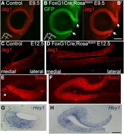
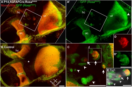
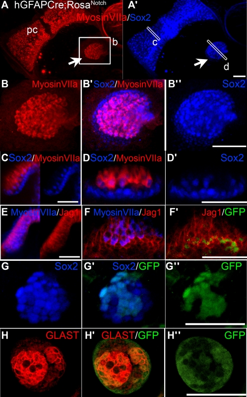
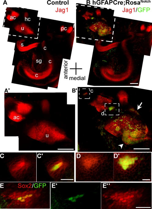
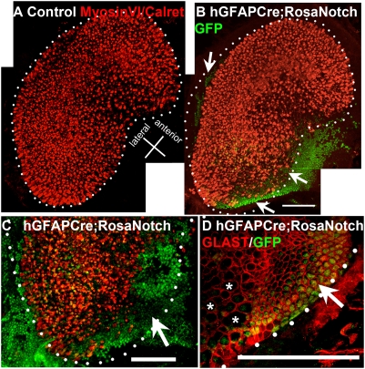
During inner ear morphogenesis, the process of prosensory specification defines the specific regions of the otic epithelium that will give rise to the six separate inner ear organs essential for hearing and balance. The mechanism of prosensory specification is not fully understood, but there is evidence that the Notch intercellular signaling pathway plays a critical role. The Notch ligand Jagged1 (Jag1) is expressed in the prosensory domains, and mutation of Jag1 impairs sensory formation. Furthermore, pharmacological inhibition of Notch in vitro during prosensory specification disrupts the prosensory process. Additionally, activation of Notch by cDNA electroporation in chick otocysts results in formation of ectopic sensory patches. Here we test whether Notch activity is sufficient for prosensory specification in the mouse, using a Cre-/loxP approach to conditionally activate the Notch pathway in nonsensory regions of the inner ear epithelia during different stages of otic vesicle morphogenesis. We find that broad ectopic activation of Notch at very early developmental stages causes induction of prosensory markers throughout the entire otic epithelium. At later stages of development, activation of Notch in nonsensory regions leads to induction of sensory patches that later differentiate to form complete ectopic sensory structures. Activation of Notch in isolated nonsensory cells results in lateral induction of Jag1 expression in neighboring cells and spreading of prosensory specification to the adjacent cells through an intercellular mechanism. These results support a model where activation of Notch and propagation through lateral induction promote prosensory character in specific regions of the developing otocyst.


Conflict of interest statement

The authors declare no conflict of interest.

Figures

Fig. 1.
Fig. 1.
Increased activation of the Notch pathway leads to failed otic vesicle closure and complete expansion of sensory domain markers. (AB’) Jag1 immunolabeling (red) in E9.5 otic vesicles from control FoxG1Cre/+ (A) and double transgenic FoxG1Cre;RosaNotch (B and B’) embryos. In control embryos, Jag1 is expressed throughout the ventral otic vesicle but is absent from the dorsal side (A). The FoxG1Cre;RosaNotch otic vesicle displays incomplete closure and broad intense labeling of Jag1 (B’, arrows). Expression of GFP (B, green), indicates transgenic Notch activation. (CH) Cochlear epithelia sections from control FoxG1Cre/+ (C, E, and G) embryos and similar sections through otic epithelia of FoxG1Cre;RosaNotch (D, F, and H) embryos at E12.5. Jag1 immunolabeling is restricted to prosensory domains in control embryos (C) and is ectopically expanded throughout the entire externalized otic epithelia in FoxG1Cre;RosaNotch embryos (D). (E and F) Similarly, Sox2 immunolabeling is restricted to the floor of the cochlear duct in control embryos (E) and expanded throughout the otic epithelium in bigenic embryos (F). Sox2 also labels delaminated neuroblasts (E, asterisk), which are reduced in FoxG1Cre;RosaNotch embryos (F). (G and H) The Notch effector Hey1 is restricted to the prosensory domains in control (G) and is expressed throughout the entire otic epithelium in bigenic embryos (H). [Scale bars, 100 μm (B) and 50 μm (H)].
Fig. 2.
Fig. 2.
Activation of Notch leads to ectopic patches of hair cells in nonsensory regions. Ectopic clusters of MyosinVIIa-labeled hair cells form in nonsensory regions of the vestibular epithelia of hGFAPCre;RosaNotch mice. Confocal projection views of vestibular epithelia from P14 hGFAPCre;RosaNotch (A and A’) or hGFAPCre/+ control (B) mice, including the utricle (ut), horizontal crista (hc), and anterior crista (ac) immunolabeled with anti-GFP (green) and the hair cell-specific marker anti-MyosinVIIa (red). In the hGFAPCre;RosaNotch epithelium, regions of ectopic hair cell formation are present between the utricle and cristae. In control vestibule, the same region (B, asterisk) is always nonsensory and devoid of hair cells. (C) High-magnification view of the boxed region in A, which contains multiple clusters of GFP+ cells (arrowheads mark examples), three of which contain ectopic MyosinVIIa+ hair cells (arrows and box). (D and D’) Split-channel views of the boxed region in C. (E) Tuj1 immunolabeling (white) shows vestibular neurites projecting to the ectopic patches in the upper region of C, as shown as a z series in Movie S2. [Scale bars, 100 μm (B, C, and E) and 50 μm (D’).]
Fig. 3.
Fig. 3.
Induced ectopic sensory patches in hGFAPCre;RosaNotch mice resemble small vestibular organs and contain hair cells and supporting cells. Examples of ectopic clusters of sensory cells from several different juvenile hGFAPCre;RosaNotch mice are shown. (A and A’) Projection views of an ectopic sensory patch (arrow) located near the posterior crista (pc) labeled with antibodies to MyosinVIIa (red) and the sensory marker Sox2 (blue). (BB’’) Higher-magnification views of the ectopic patch in A and A’. (C) Transverse view through the boxed region of the posterior crista shown in A and A’ showing the merged Sox2/MyosinVIIa labeling (Left), as well as the Sox2 labeling alone (Right). (D and D’) Transverse views through the boxed region of the ectopic sensory patch shown in A and A’. (E) Transverse view of a crista labeled with anti-MyosinVIIa (blue) and anti-Jag1 (red), showing the merged channels (Left), as well as the Jag1 alone (Right). (FF’’) Transverse view of an ectopic sensory cluster labeled with anti-MyosinVIIa (blue), anti-Jag1 (red), and anti-GFP (green). (GG’’) An ectopic cluster colabeled with anti-GFP (green) and anti-Sox2 (blue). (HH’’) An ectopic cluster colabeled with antibodies to GFP (green) and the mature supporting cell marker GLAST (red). A, A’, and BB’’ are brightest point projections; all others are single optical sections. (Scale bars, 100 μm.)
Fig. 4.
Fig. 4.
Activation of Notch during prosensory specification leads to up-regulation of Jag1 and Sox2 in cis and trans. (A) Jag1 immunolabeling (red) in control E13.5 otic capsules is present in the five vestibular organs as well as the cochlea. (A’) An enlarged view of the boxed region in A showing the Jag1 labeling in the utricle (u) and anterior crista (ac). (B) An E13.5 hGFAPCre;RosaNotch otic capsule immunolabeled for Jag1 (red) and GFP (green, indicating transgenic NotchIC expression). (B’) An enlarged view of the boxed region in B showing the anterior crista and utricle labeled for Jag1 (red) and GFP (green). The pattern of Jag1 labeling in the hGFAPCre;RosaNotch utricle is expanded in a medial posterior protruding region (arrowhead; compare B’ with A’) of GFP+ cells. There are ectopic clusters of Jag1 labeling lateral to the utricle (arrow) and on either side of the anterior crista (boxed regions in B’), all of which contain GFP+ cells. A–A’ and B–B’ are montages of images compiled from brightest point projections of multiphoton excitation micrographs. (C and C’) A single optical section taken from the upper boxed region in B shows two adjacent GFP+ cells brightly labeled for Jag1, and neighboring, GFP−, cells also show up-regulated Jag1 expression. (D and D’) An enlarged view of a single optical section taken from the lower boxed region in B’ shows a cluster of about 15 GFP+ cells that have marked up-regulation of Jag1 expression, whereas surrounding GFP− cells also show up-regulation of Jag1 (D). (E and E’) Another hGFAPCre;RosaNotch sample displaying an ectopic Sox2+ cluster of cells between the utricle and anterior crista. This cluster consists of four GFP+ cells (E’) that are brightly labeled for Sox2 and are surrounded by a perimeter of ectopic Sox2+, GFP−, cells (EE’’). ac, anterior crista; c, cochlea; hc, horizontal crista; pc, posterior crista; s, saccule; sg, spiral ganglia; u, utricle. [Scale bars, 100 μm (A’, B, and B’) and 20 μm (C’, D’, and E’’).]
Fig. 5.
Fig. 5.
Notch activation inhibits hair cell formation in vestibular organs. Regions of high-GFP+ cell density in utricles of hGFAPCre;RosaNotch mice contain fewer hair cells and more supporting cells. (A) A montage view of a normal control utricle, stained with an antibody mixture to MyosinVI and Calretinin (both in red) to label hair cells and their associated afferent neurites, displays dense patterning of hair cells within a clearly defined border (dotted line) outlining its characteristic kidney shape. (B) An hGFAPCre;RosaNotch utricle, shown as a montage, displays markedly reduced MyosinVI/Calretinin labeling (red) in areas with a high density of GFP+ cells (arrows, green) and a reduced overall hair cell area lacking a clear border in places. (C) A higher-magnification view of the posterior region of another hGFAPCre;RosaNotch utricle labeled as above shows a large GFP+ region devoid of hair cells (arrow). (D) In an hGFAPCre;RosaNotch utricle, GLAST immunolabeling (specific to supporting cell membranes) displays an abnormally continuous pattern in regions of high-GFP+ cell density, whereas nearby areas with fewer GFP+ cells display the normal pattern of GLAST distribution, where voids in expression of this marker occur in areas containing hair cells (asterisks). The dotted lines in BD represent what would be the normal border region of the utricle as shown in A. (Scale bars, 100 μm.)

Similar articles

Cited by

References

    1. Adam J, et al. Cell fate choices and the expression of Notch, Delta and Serrate homologues in the chick inner ear: Parallels with Drosophila sense-organ development. Development. 1998;125:4645–4654. - PubMed
    1. Lewis AK, Frantz GD, Carpenter DA, de Sauvage FJ, Gao WQ. Distinct expression patterns of Notch family receptors and ligands during development of the mammalian inner ear. Mech Dev. 1998;78:159–163. - PubMed
    1. Morrison A, Hodgetts C, Gossler A, Hrabé de Angelis M, Lewis J. Expression of Delta1 and Serrate1 (Jagged1) in the mouse inner ear. Mech Dev. 1999;84:169–172. - PubMed
    1. Murata J, Tokunaga A, Okano H, Kubo T. Mapping of Notch activation during cochlear development in mice: Implications for determination of prosensory domain and cell fate diversification. J Comp Neurol. 2006;497:502–518. - PubMed
    1. Hayashi T, et al. Hesr1 and Hesr2 may act as early effectors of Notch signaling in the developing cochlea. Dev Biol. 2008;316:87–99. - PMC - PubMed

Publication types

Substances

LinkOut - more resources