. 2010 Oct 1;330(6000):94-7.
doi: 10.1126/science.1192280. Epub 2010 Aug 26.

Mitotic recombination in patients with ichthyosis causes reversion of dominant mutations in KRT10

Affiliations

Mitotic recombination in patients with ichthyosis causes reversion of dominant mutations in KRT10

Keith A Choate et al. Science. .

Abstract

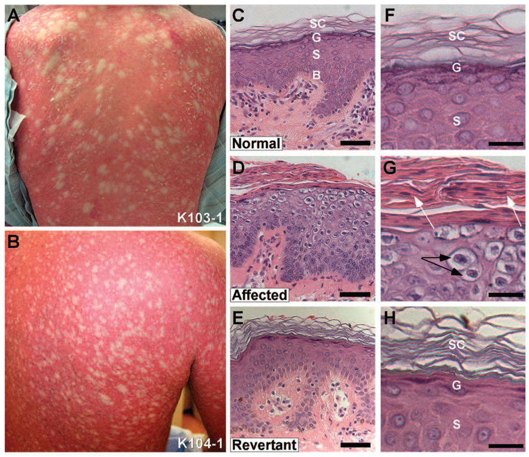
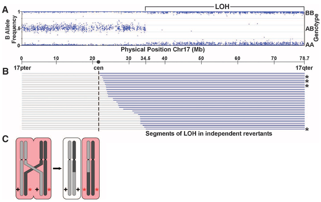
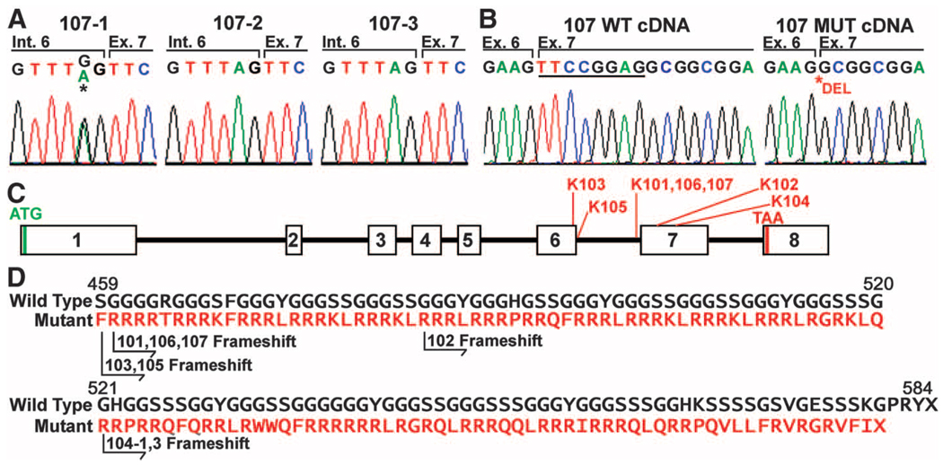
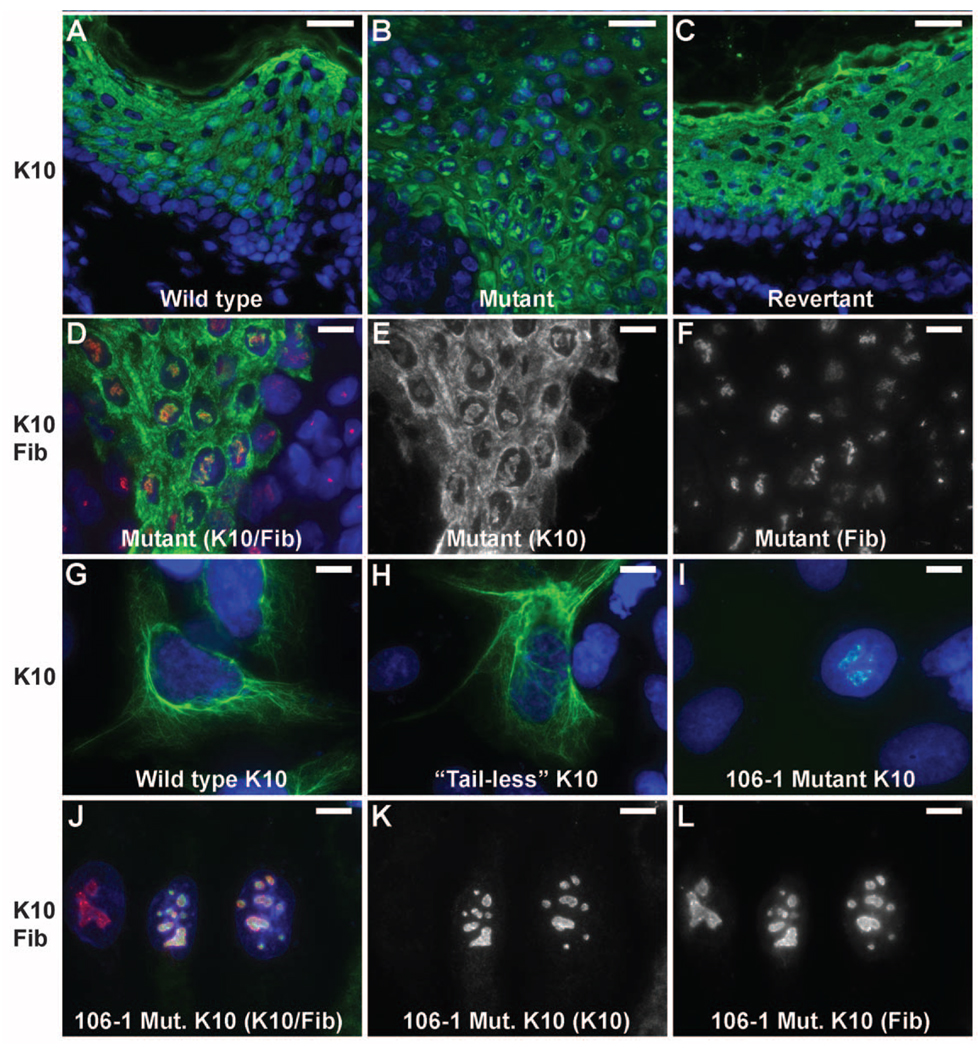
Somatic loss of wild-type alleles can produce disease traits such as neoplasia. Conversely, somatic loss of disease-causing mutations can revert phenotypes; however, these events are infrequently observed. Here we show that ichthyosis with confetti, a severe, sporadic skin disease in humans, is associated with thousands of revertant clones of normal skin that arise from loss of heterozygosity on chromosome 17q via mitotic recombination. This allowed us to map and identify disease-causing mutations in the gene encoding keratin 10 (KRT10); all result in frameshifts into the same alternative reading frame, producing an arginine-rich C-terminal peptide that redirects keratin 10 from the cytokeratin filament network to the nucleolus. The high frequency of somatic reversion in ichthyosis with confetti suggests that revertant stem cell clones are under strong positive selection and/or that the rate of mitotic recombination is elevated in individuals with this disorder.


Figures

Fig. 1
Fig. 1
Frequent revertants in Ichthyosis with confetti. (A, B) The backs of an 18 year-old female subject (103-1) and 42 year-old male (104-1) show background redness and scaling with hundreds of white, normal-appearing "confetti" spots. (C) Histology of normal human skin showing basal layer (labeled ‘B’), stratum spinosum (‘S’), granular layer (‘G’) and stratum corneum (‘SC’). (D) Affected skin shows loss of differentiation of all layers above the basal layer and hypercellularity with increased epidermal thickness. There is no granular layer, marked peri-nuclear vacuolization in the suprabasal epidermis, and retention of cell nuclei in the stratum corneum. (E) Revertant skin shows normalization of epidermal thickness and architecture, with normal granular layer, normal spinous layer, and stratum corneum. (Scale bars in C–E = 50 µm) (F) High power of spinous layer in normal epidermis with intercellular spines visible, overlying granular layer with purple keratohyalin granules and basket weave stratum corneum. (G) High power view of affected skin shows peri-nuclear vacuolization (black arrows), lack of keratohyalin granules, and retained nuclei (white arrows) in the stratum corneum. (H) High power view of revertant skin shows normal spinous layer with intercellular spines, granular layer with purple keratohyalin granules in keratinocytes, and basket weave stratum corneum. (Scale bars in F–H = 25 µm)
Fig. 2
Fig. 2
Revertant spots show loss of heterozygosity on 17q. (A) Genotypes on chromosome 17 from revertant keratinocytes of IWC subject 106-1 are shown. From 17pter to 34.5M base pairs, genotypes show the expected heterozygosity with genotypes identical to blood and disease keratinocyte DNA (Fig. S2), while from 34.5M base pairs to 17qter, genotypes are homozygous with no change in diploid copy number (Fig. S2). (B) Results of genotyping keratinocytes of 32 revertant spots from 7 unrelated IEC subjects (106-1 revertants denoted with *). Gray lines = genomic segments with heterozygous genotypes matching blood DNA; blue lines = segments showing copy-neutral LOH. These results are consistent with IWC being caused by a dominant allele distal to 34.5M bp that is lost by mitotic recombination, as depicted in panel (C).
Fig. 3
Fig. 3
Mutations in KRT10 (keratin 10) cause IWC. (A) Sanger sequencing confirms de novo mutation in KRT10 from Illumina sequencing that is absent in the parents (107-2 and 107-3) but present in the affected offspring (107-1) that abolishes the splice acceptor site of intron 6. (B) Abnormal splicing of KRT10. cDNA from diseased keratinocytes of 107-1 shows 2 splice forms, one wild-type (WT) and one using an AG splice acceptor that deletes 8 bases from WT cDNA (underlined). (C) The genomic structure of KRT10 is shown. The locations of mutations found in IWC kindreds are indicated. (D) IWC frameshifts all produce an arginine-rich C-terminal peptide. The normal sequence of the C-terminal 226 amino acids of keratin 10 is shown; below, in red, the sequence of the frameshift peptides found in IWC are shown, with the position of the frameshift in each kindred indicated.
Fig. 4
Fig. 4
Mutant keratin 10 is redirected to nucleoli in vivo and in vitro. (A–C) Images of normal, mutant and revertant skin stained with DAPI and antibodies to keratin 10 reveal discrete foci of nuclear keratin 10 in mutant skin which are absent in normal and revertant skin. (Scale bars = 50 µm). (D–F) Co-staining with the nucleolar marker fibrillarin shows that keratin 10 is in the nucleolus. (Scale bars = 10 µm) (G–I) Constructs bearing wild-type keratin 10 (G), keratin 10 truncated at the beginning of the tail domain (amino acid 459) (H) and keratin 10 with the kindred 106 frameshift mutation beginning at codon 460 (I) were expressed in PLC cells and stained with DAPI and monoclonal antibody to keratin 10. Wild-type and ‘tailless’ K10 integrate into the cytoplasmic filament network while the IWC mutant K10 localizes to nucleoli as shown by costaining with fibrillarin (J–L). (Scale bars in G–L = 10 µm).

Comment in

References

    1. Camenzind M, Harms M, Chavaz O, Saurat JH. Ichythose en confettis. Ann. Dermatol. Venereol. 1984;111:675. - PubMed
    1. Brusasco A, et al. A case of congenital reticular ichthyosiform erythroderma – ichthyosis ‘en confetis’. Dermatology. 1994;188:40. - PubMed
    1. Marghescu S, Anton-Lamprecht I, Rudolph PO, Kaste R. Congenital reticular ichthyosiform erythroderma. Hautarzt. 1984;35:522. - PubMed
    1. Information on materials and methods is available on Science Online.

    1. UCSC genome browser, build hg18 of the human genome, http://genome.ucsc.edu/cgi-bin/hgGateway, of the human genome.

Publication types

MeSH terms

LinkOut - more resources