. 2010 Aug;22(8):2594-617.
doi: 10.1105/tpc.109.073171. Epub 2010 Aug 26.

Phosphoenolpyruvate provision to plastids is essential for gametophyte and sporophyte development in Arabidopsis thaliana

Affiliations

Phosphoenolpyruvate provision to plastids is essential for gametophyte and sporophyte development in Arabidopsis thaliana

Veena Prabhakar et al. Plant Cell. 2010 Aug.

Abstract

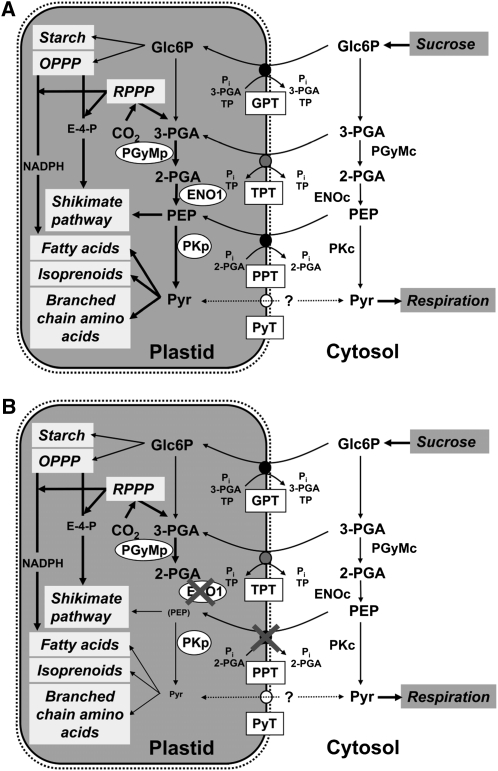
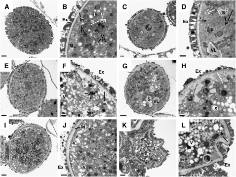
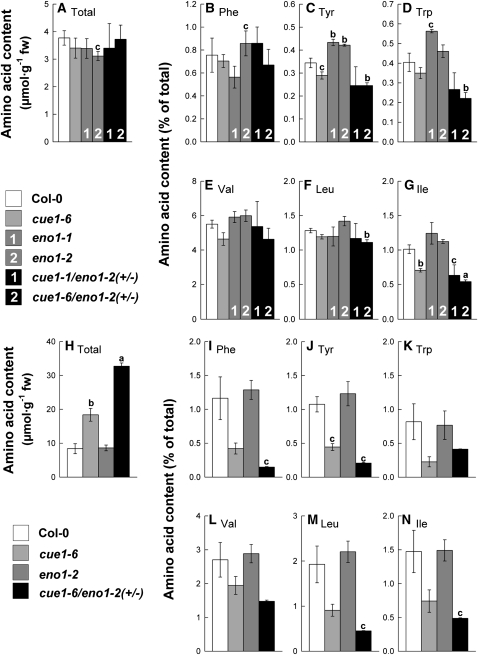
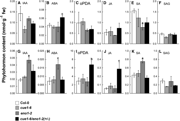
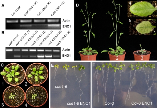
Restriction of phosphoenolpyruvate (PEP) supply to plastids causes lethality of female and male gametophytes in Arabidopsis thaliana defective in both a phosphoenolpyruvate/phosphate translocator (PPT) of the inner envelope membrane and the plastid-localized enolase (ENO1) involved in glycolytic PEP provision. Homozygous double mutants of cue1 (defective in PPT1) and eno1 could not be obtained, and homozygous cue1 heterozygous eno1 mutants [cue1/eno1(+/-)] exhibited retarded vegetative growth, disturbed flower development, and up to 80% seed abortion. The phenotypes of diminished oil in seeds, reduced flavonoids and aromatic amino acids in flowers, compromised lignin biosynthesis in stems, and aberrant exine formation in pollen indicate that cue1/eno1(+/-) disrupts multiple pathways. While diminished fatty acid biosynthesis from PEP via plastidial pyruvate kinase appears to affect seed abortion, a restriction in the shikimate pathway affects formation of sporopollonin in the tapetum and lignin in the stem. Vegetative parts of cue1/eno1(+/-) contained increased free amino acids and jasmonic acid but had normal wax biosynthesis. ENO1 overexpression in cue1 rescued the leaf and root phenotypes, restored photosynthetic capacity, and improved seed yield and oil contents. In chloroplasts, ENO1 might be the only enzyme missing for a complete plastidic glycolysis.


Figures

Figure 1.
Figure 1.
Metabolic Role of PEP in Plastids of Heterotrophic or Mixotrophic Tissues (i.e., Developing Seeds). In wild-type plants (A), PEP can be imported from the cytosol by PPT, or it may be produced from 3-PGA by the glycolytic sequence involving PGyM and ENO. Both enzymes exist as plastidic and cytosolic forms. In the stroma, PEP together with erythrose 4-phosphate (E-4-P) can enter the shikimate pathway for the biosynthesis of aromatic amino acids and derived compounds, or after conversion to pyruvate by PK, it can be fed into the biosynthesis of fatty acids, isoprenoids, or branched-chain amino acids. Pyruvate may also be imported by a pyruvate transporter (PyT). Other transporters of the phosphate translocator family, such as GPT or the triose phosphate/PT (TPT), may import Glc6P or 3-PGA, respectively. Glc6P can be fed into OPPP and starch biosynthesis. Note that TPT is not likely to be expressed in heterotrophic tissues. The OPPP produces reducing equivalents in the form of NADPH required for anabolic reactions and metabolic intermediates, such as E-4-P. In mixotrophic plastids, 3-PGA and reducing equivalents can be produced by the Calvin cycle (reductive pentose phosphate pathway [RPPP]). By cytosolic glycolysis, imported sucrose can be metabolized to pyruvate, which is subjected to respiration in the mitochondria. In (B), the consequences of a deficiency in both PPT1 and ENO1 are shown. Most likely all metabolic pathways shaded in light gray within the plastids would be negatively affected, which would also feed back on processes taking place in the cytosol.
Figure 2.
Figure 2.
Phenotype of Heterozygous eno1 Mutants in the Homozygous cue1 Background (ccEe) Compared with Wild-Type and cue1 Plants Grown for 8 Weeks in the Greenhouse. (A) Comparison of the growth phenotype of Col-0 (1), eno1-2 (2), cue1-1 (3), and cue1-1/eno1-2 (4) plants. The inset shows a detailed view of cue1-1/eno1-2(+/−) (5). (B) Opened bud of an early (stage 10) wild-type (Col-0) flower. (C) Opened bud of an early (stage 10) flower of cue1-1/eno1-2(+/−) with degenerated stamens. (D) Flower and silique development of cue1-6. (E) Schematic representation of the position of flowers and siliques shown in (D) and (F) (F) Flower and silique development of cue1-6/eno1-2(+/−) plants. (G) Destained mature siliques of Col-0 (1), pOCA (2), cue1-1 (3), cue1-6/eno1-2(+/−) (4), and cue1-1/eno1-2(+/−) (5) Developmental stages of flowers and siliques shown in (D) and (F) are based on the position of the flowers/siliques at the raceme starting from the topmost to lower positions. The numbers in (E) represent the positions of flowers and/or siliques shown in (D) and (F). Bars = 5 mm in (D) and (F).
Figure 3.
Figure 3.
Phenotypes of Ovules and Mature Seeds of Heterozygous eno1 Mutants in the Homozygous cue1 Background (ccEe) Compared with Wild-Type or cue1 Plants. (A) Ovule of a wild-type (Col-0) plant. (B) Ovule of cue1-1/eno1-2(+/−) with a wild-type-like appearance. (C) Ovule of cue1-1/eno1-2(+/−) with a swollen embryo sac. (D) Ovule of cue1-1/eno1-2(+/−) lacking an embryo sac. (E) Degenerated ovule of cue1-1/eno1-2(+/−). (F) Wild-type (pOCA) seeds. (G) Seeds of cue1-1. (H) Phenotype of seeds of cue1-1/eno1-2(+/−) showing either a wild-type-like appearance (class I seeds; 1) or that were intermediately and strongly reduced in size (class II, 2; and class III seeds, 3). Bar = 50 μm in (A) to (E). [See online article for color version of this figure.]
Figure 4.
Figure 4.
Cross Sections of Pollen Sacs as well as Vitality (Alexander) and DAPI Staining of Pollen from Heterozygous eno1 Mutants in the Homozygous cue1 Background (ccEe) Compared with the Wild Type. (A) Cross section of a wild-type (Col-0) pollen sac. (B) Cross section of a pollen sac of cue1-1/eno1-2(+/−). (C) Cross section of a pollen sac from cue1-6/eno1-2(+/−). (D) Vitality staining of wild-type pollen (pOCA). (E) Vitality staining of pollen from cue1-1/eno1-2(+/−) with an example of a viable (1) and an aborted (2) pollen. (F) Bright-field image of wild-type pollen (pOCA). (G) DAPI staining of the wild-type pollen shown in (F). (H) Bright-field image of wild-type-like pollen and degenerated pollen (arrow) of cue1-1/eno1-2(+/−). (I) DAPI staining of the pollen grains shown in (H). The degenerated pollen is marked by an arrow. [See online article for color version of this figure.]
Figure 5.
Figure 5.
Cross Sections of Differently Affected Pollen Grains of Heterozygous eno1 Mutants in the Homozygous cue1 Background (ccEe) Analyzed by Transmission Electron Microscopy in the Tricellular Stage in Comparison to Wild-Type Col-0. (A) Cross section of a wild-type (Col-0) pollen grain. (B) Close-up of the wild-type pollen grain shown in (A). (C) to (L) Phenotypic changes in the ultrastructure of pollen grains observed in pollen sacs of ccEe plants. (C) Pollen grain of cue1-1/eno1-2(+/−) with a wild-type-like appearance. (D) Close-up of the pollen grain shown in (C) with increased numbers of starch granules in the plastids and a slightly deformed and swollen intine. (E) Pollen grain of cue1-1/eno1-2(+/−) with a wild-type-like size but affected exine and intine structures. (F) Close-up of the pollen grain shown in (E) with a focus on the underdeveloped exine structure and the strongly deformed intine. (G) Pollen grain of cue1-1/eno1-2(+/−) with a deformed pollen wall, high numbers of starch granules, and large vacuole-like structures. (H) Close-up of the pollen grain shown in (G) with a focus on the impaired exine and intine structures. (I) Pollen grain of cue1-6/eno1-2(+/−) with a wild-type-like appearance but an impaired exine structure. (J) Close-up of the pollen grain shown in (I) with a focus on the underdeveloped exine structure. (K) Strongly deformed pollen grain of cue1-6/eno1-2(+/−). (L) Close-up of the pollen grain shown in (K). Ex, exine; In, intine; L, lipid body; ER, endoplasmatic reticulum; V, vacuoles; VL, vacuole-like bodies; M, mitochondria; P, plastids; S, starch granules. Bars = 2 and 0.5 μm for the overviews and close-ups, respectively.
Figure 6.
Figure 6.
Contents of Selected Amino Acids Extracted from Flower Buds or Rosette Leaves of the Wild Type (Col-0), cue1-6 and eno1 Single Mutant, and the Heterozygous eno1 Mutants in the Homozygous cue1 Background (ccEe). Flower buds ([A] to [G]) and rosette leaves ([H] to [N]). The data represent the mean ± se of n = 5 ([A] to [G]) or n = 3 ([H] to [N]) independent experiments. Statistical significance of differences between the parameters were assessed by the Welch test with probability values of P < 0.001 (a), P < 0.01 (b), and P < 0.05 (c) indicated above the respective bars. Contents of total soluble amino acid ([A] and [H]) were estimated from the sum of all recognized proteinogenic amino acid after separation by HPLC. The relative contents of the aromatic amino acids Phe, Tyr, Trp ([B] to [D] and [I] to [K]) and the branched-chain amino acids Val, Leu, and Ile ([E] to [G] and [L] to [N]) were expressed as a percentage fraction of the total amino acid content in flower buds (A) and rosette leaves (H), respectively.
Figure 7.
Figure 7.
Contents of Phytohormones in Flowers or Rosette Leaves of Col-0 Wild Type, cue1-6 and eno1-2 Single Mutants, and the Heterozygous eno1-2 Mutant in the Homozygous cue1-6 Background (cue1-6/eno1-2[+/−]). Flowers ([A] to [F]) and rosette leaves ([G] to [L]). Phytohormones identified by liquid chromatography–mass spectrometry separation were IAA ([A] and [G]), ABA ([B] and [H]), oPDA ([C] and [I]), JA ([D] and [J]), SA ([E] and [K]), and SAG ([F] and [L]). The data represent the mean ± se of n = 3 independent experiments. Statistical significance of differences between the parameters was assessed by the Welch test with probability values of P < 0.01 (a), P < 0.02 (b), and P < 0.05 (c) indicated above the respective bars.
Figure 8.
Figure 8.
Ectopic Overexpression of ENO1 Rescues the cue1 Phenotype. (A) RT-PCR with RNA extracted from leaves of wild-type (Col-0) or transformants overexpressing ENO1 in the Col-0 background. (B) RT-PCR with RNA extracted from leaves of cue1-6 or transformants overexpressing ENO1 in the cue1-6 background. (C) Rosette phenotypes of the wild type (1), an ENO1-overexpressing line [cue1-6 ENO1 (4)] with a high transcript abundance of ENO1 (2), the cue1-6 mutant (3), and an ENO1-overexpressing line [cue1-6 ENO1 (1)] with a low transcript abundance of ENO1 (4). (D) Growth phenotype of flowering Col-0 wild-type (1), an ENO1-overexpressing line in the cue1-6 background [cue1-6 ENO1 (4)] with a high transcript abundance of ENO1 (2), and cue1-6 (3) plants. The inset shows the rescue of the leaf phenotype of cue1-6 overexpressing ENO1 [line cue1-6 ENO1 (4)] in comparison with cue1-6 (5). (E) Rescue of the retarded root phenotype of the cue1-6 mutant by overexpression of ENO1 in the cue1-6 background. Three cue1-6 mutant plants are compared with one ENO1-overexpressing line [i.e., cue1-6 ENO1 (4)]. (F) Phenotypic comparison of wild-type Col-0 with Col-0 plants overexpressing ENO1.
Figure 9.
Figure 9.
Light Saturation Curves of Photosynthetic ETRs Determined by Imaging Pulse Amplitude Modulation Fluorometry. (A) Comparison of light saturation curves between Col-0 (open circles), eno1-2 (triangles), Col-0 ENO1 (A) (closed circles), and Col-0 ENO1 (C) (squares). (B) Comparison of light saturation curves between Col-0 (cirlces), cue1-6 (squares), and cue1-6 ENO1 (4) (triangles). The data represent the mean ± se of n = 9 measurements on individual plants per line.

Similar articles

Cited by

References

    1. Aarts M.G., Keijzer C.J., Stiekema W.J., Pereira A. (1995). Molecular characterization of the CER1 gene of arabidopsis Arabidopsis involved in epicuticular wax biosynthesis and pollen fertility. Plant Cell 7: 2115–2127 - PMC - PubMed
    1. Aharoni A., Dixit S., Jetter R., Thoenes E., van Arkel G., Pereira A. (2004). The SHINE clade of AP2 domain transcription factors activates wax biosynthesis, alters cuticle properties, and confers drought tolerance when overexpressed in Arabidopsis. Plant Cell 16: 2463–2480 - PMC - PubMed
    1. Alexander M.P. (1969). Differential staining of aborted and nonaborted pollen. Stain Technol. 44: 117–122 - PubMed
    1. Andre C., Froehlich J.E., Moll M.R., Benning C. (2007). A heteromeric plastidic pyruvate kinase complex involved in seed oil biosynthesis in Arabidopsis. Plant Cell 19: 2006–2022 - PMC - PubMed
    1. Ariizumi T., Hatakeyama K., Hinata K., Inatsugi R., Nishida I., Sato S., Kato T., Tabata S., Toriyama K. (2004). Disruption of the novel plant protein NEF1 affects lipid accumulation in plastids of the tapetum and exine formation of pollen, resulting in male sterility in Arabidopsis thaliana. Plant J. 39: 170–181 - PubMed

Publication types

MeSH terms

Substances

LinkOut - more resources