. 2010 Aug;22(8):2781-95.
doi: 10.1105/tpc.110.076778. Epub 2010 Aug 26.

Tobacco GTBP1, a homolog of human heterogeneous nuclear ribonucleoprotein, protects telomeres from aberrant homologous recombination

Affiliations

Tobacco GTBP1, a homolog of human heterogeneous nuclear ribonucleoprotein, protects telomeres from aberrant homologous recombination

Yong Woo Lee et al. Plant Cell. 2010 Aug.

Abstract

Telomeres are nucleoprotein complexes essential for the integrity of eukaryotic chromosomes. Cellular roles of single-stranded telomeric DNA binding proteins have been extensively described in yeast and animals, but our knowledge about plant single-strand telomeric factors is rudimentary. Here, we investigated Nicotiana tabacum G-strand-specific single-stranded telomere binding proteins (GTBPs), homologs of a human heterogeneous nuclear ribonucleoprotein. GTBPs bound specifically to the plant single-stranded (TTTAGGG)(4) telomeric repeat element in vitro and were associated with telomeric sequences in tobacco BY-2 suspension cells. Transgenic plants (35S:RNAi-GTBP1), in which GTBP1 was suppressed, exhibited severe developmental anomalies. In addition, the chromosomes of 35S:RNAi-GTBP1 cells displayed elongated telomeres, frequent formation of extrachromosomal telomeric circles, and numerous abnormal anaphase bridges, indicating that GTBP1 knockdown tobacco plants experienced genome instability. GTBP1 inhibited strand invasion, an initial step in interchromosomal homologous recombination. We propose that GTBP1 plays a critical role in telomere structure and function by preventing aberrant interchromosomal telomeric homologous recombination in tobacco.


Figures
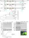
Figure 1.
Figure 1.
Structure and Expression of Nt GTBPs. (A) Schematic representation of three tobacco GTBP cDNAs and their predicted proteins. The solid lines show 5′- and 3′-untranslated regions. The bars depict coding regions. The RRMs (RRM1 and RRM2) are indicated. PCR primers for partial cDNA cloning, 5′- and 3′-RACE, and RT-PCR are indicated by arrows with triangles, circles, and squares, respectively. (B) Phylogenetic relationship of 10 GTBP homologs from tobacco, Arabidopsis, rice, and human. The tree was constructed using MEGA4 software with the neighbor-joining method and the summed branch length of the tree is 3.21911554. Amino acid identities of these proteins to GTBP1 are shown in right side. A text file of the alignment used to generate this tree is available as Supplemental Data Set 1 online. (C) Expression profiles of three GTBP paralogs. Total RNA was isolated from BY-2 cells over the course of 9 d (left gel) and from leaves, stems, and roots of 1-month-old tobacco plants (right gel) and analyzed by RT-PCR using gene-specific primers. Growth curve of BY-2 cells is shown in the bottom of left panel. Tobacco leaves that are in four different developmental stages are shown in the bottom of the right panel. Ng TRF1 and Nt cyM were used as G-phase and M-phase markers, respectively. The level of elongation factor 1α (EF1α) transcript is shown as a loading control. [See online article for color version of this figure.]
Figure 2.
Figure 2.
Nt GTBPs Bind Plant Single-Stranded Telomeric Repeats with Sequence Specificity. (A) GTBPs bind plant single-stranded telomeric sequences. Various quantities (0, 0.1, 0.3, and 0.9 μg) of three Nt GTBP paralogs were incubated with radiolabeled single-stranded (TTTAGGG)4 (PT4) repeats and analyzed in a gel retardation assay. (B) Sequence-specific binding activity of GTBPs. Nt GTBPs (1 μg) were incubated with radiolabeled PT4 in the presence of a 10- or 50-fold excess of unlabeled plant (PT4), human (HT4), C. elegans (CT4), or nonspecific (NS) telomeric DNA as competitors. The first “0” lane of each gel contained radiolabeled PT4 without protein. (C) Gel retardation assays with various telomere repeats. GTBPs (1 μg) were analyzed with radiolabeled PT2, PT3, or PT4 (containing two, three, or four repeats, respectively). (D) Gel retardation assays with a series of single nucleotide mutants of PT4. Each mutant (M1 to M7) contained a single nucleotide substitution in the repeated sequence, as listed below the gels.
Figure 3.
Figure 3.
GTBP1 Is Associated with Telomeric Sequences in Tobacco BY-2 Cells. (A) Nuclear localization of HA-GTBP1. 35S:HA-GTBP1 was transformed into BY-2 cells, and localization of HA-GTBP1 was examined by immunohistochemistry using an anti-HA antibody. The BY-2 cells were viewed by fluorescence microscopy under dark field and light field. The nuclei were stained with DAPI. Bars = 10 μm. (B) ChIP assay. The genomic DNA-protein complex from wild-type and 35S:HA-GTBP1 BY-2 cells was fragmented by sonication and subjected to immunoprecipitation using an anti-HA antibody. The coimmunoprecipitated DNA was hybridized with 32P-labeled (TTTAGGG)70 or HRS60 repeated tobacco DNA sequences.
Figure 4.
Figure 4.
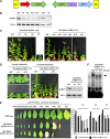
Construction and Characterization of 35S:RNAi-GTBP1 Knockdown Transgenic Tobacco Plants. (A) Schematic structure of GTBP1 RNAi binary vector construct. The 35S:RNAi-GTBP1 vector includes the inverted-repeat sequence of the N-terminal (122 to 498 bp) or C-terminal (726 to 1070 bp) regions of GTBP1 cDNA. LB, left border; OCS ter, octopine synthase terminator; NPTII, neomycin phosphotransferase II; RB, right border. (B) Repression of GTBP1 mRNA in transgenic plants. Total leaf RNA from wild-type and independent T0 35S:RNAi-GTBP1 lines was analyzed by RT-PCR. EF1α was used as a loading control. (C) Gross morphology of 2-month-old wild-type and independent T0 transgenic lines grown under greenhouse conditions. (D) Morphology of 3-week-old and 2-month-old wild-type and T1 RNAi transgenic lines (#1 and #13). GTBP1 transcript levels were determined by RT-PCR in T1 generation (shown at the right with EF1α as a control). (E) Morphological comparison of leaves from 2-month-old wild-type and T1 transgenic (#1 and #13) plants. Leaf number and stem length of wild-type and 35S:RNAi-GTBP1 (T0 and T1) tobacco plants are shown in the right panel. Bar = 3 cm. (F) Single-strand telomere binding activities of wild-type and T1 RNAi transgenic seedlings. The cell-free nuclear extracts containing 10 μg of protein were prepared from wild-type and T1 transgenic seedlings (#1 and #13), incubated with PT4, and analyzed by gel retardation assays as described in Figure 2. Arrow indicates protein-PT4 complex. [See online article for color version of this figure.]
Figure 5.
Figure 5.
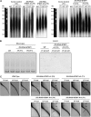
Telomere Deregulation in 35S:RNAi-GTBP1 Tobacco Plants. (A) Telomere elongation in T0 and T1 transgenic plants. Genomic DNA was purified from the 27th leaves of wild-type plants and the 7th, 15th, and 27th leaves of T0 and T1 transgenic (#1 and #13) plants. After digestion with TaqI, DNA was subjected to TRF analysis by pulse-field gel electrophoresis. (B) Telomerase activity in wild-type and T1 transgenic (#1 and #13) plants. The level of telomerase activity was examined in shoot apexes and various leaves by PCR-based TRAP assays. (C) Formation of extrachromosomal t-circles in T0 and T1 transgenic (#1 and #13) plants. Genomic DNA from the 7th, 15th, and 27th leaves of wild-type and T0 and T1 35S:RNAi-GTBP1 (lines #1 and #13) tobacco plants were subjected to TRF analysis by 2D pulse-field gel electrophoresis. Linear telomeric DNA and extrachromosomal t-circles are indicated by arrows and arrowheads, respectively.
Figure 6.
Figure 6.
Formation of Anaphase Bridges in T1 35S:RNAi-GTBP1 Transgenic Cells. (A) Anaphase chromosomes from wild-type and T1 35S:RNAi-GTBP1 (lines #1 and #13) anther cells. Anaphase chromosome spreads were obtained from anthers of wild-type and 35S:RNAi-GTBP1 transgenic lines (#1 and #13), stained with DAPI, and observed by fluorescence microscopy. Abnormal anaphase bridges were detected in transgenic meiotic cells. Frequencies of single, double, and multiple anaphase bridges are indicated in the right panel. Bars = 10 μm. (B) FISH analysis of anaphase chromosomes in wild-type and T1 35S:RNAi-GTBP1 (lines #1 and #13) meiotic cells using a (TTTAGGG)70 repeat telomeric fluorescent probe. Chromosomal DNA was denatured and incubated with Texas red-dUTP–incorporated (TTTAGGG)70 telomeric probe. The chromosomes were counterstained with DAPI and observed using fluorescence microscopy. Arrows indicate telomeric signals at the fusion points of anaphase bridges. Percentages of anaphase bridges with telomeric sequences detected by FISH are indicated in the right panel. Bars = 10 μm.
Figure 7.
Figure 7.
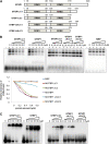
Nt GTBP1 Inhibits Telomeric Strand Invasion in Vitro. (A) Schematic representation of full-length and deletion mutants of GTBP1. (B) Strand invasion assay. Increasing concentrations of GTBP1 derivatives (0, 0.2, 0.4, 0.6, and 0.8 μM) were incubated with a 32P-labeled single-stranded (TTTAGGG)8 repeat probe with T-vector plasmid containing double-stranded (TTTAGGG)70 telomere repeats. The degree of invasion of the single-stranded telomeric probe into the plasmid was determined by the shifted band intensity. The first “−” lane of each gel contained radiolabeled PT8 without protein. The graph in the bottom panel shows relative intensities of shifted bands. (C) Binding activities of full-length and deletion mutants of GTBP1 to single-strand telomere sequences. Various concentrations (0, 0.2, 0.4, 0.6, and 0.8 μM) of GTBP1 derivatives were analyzed with radiolabeled single-stranded (TTTAGGG)8 (PT8) repeats. [See online article for color version of this figure.]

Similar articles

Cited by

References

    1. Amiard S., et al. (2007). A topological mechanism for TRF2-enhanced strand invasion. Nat. Struct. Mol. Biol. 14: 147–154 - PubMed
    1. Bae H., Choi S.M., Yang S.W., Pai H.S., Kim W.T. (2009). Suppression of the ER-localized AAA ATPase NgCDC48 inhibits tobacco growth and development. Mol. Cells 28: 57–65 - PubMed
    1. Baumann P., Cech T.R. (2001). Pot1, the putative telomere end-binding protein in fission yeast and humans. Science 292: 1171–1175 - PubMed
    1. Bilaud T., Brun C., Ancelin K., Koering C.E., Laroche T., Gilson E. (1997). Telomeric localization of TRF2, a novel human telobox protein. Nat. Genet. 17: 236–239 - PubMed
    1. Blackburn E.H. (1991). Structure and function of telomeres. Nature 350: 569–573 - PubMed

Publication types

MeSH terms

LinkOut - more resources