. 2010 Nov;30(11):2301-9.
doi: 10.1161/ATVBAHA.110.209460. Epub 2010 Aug 26.

Cardiovascular pathology in Hutchinson-Gilford progeria: correlation with the vascular pathology of aging

Affiliations

Cardiovascular pathology in Hutchinson-Gilford progeria: correlation with the vascular pathology of aging

Michelle Olive et al. Arterioscler Thromb Vasc Biol. 2010 Nov.

Abstract

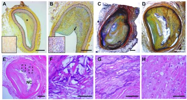
Objective: Children with Hutchinson-Gilford progeria syndrome (HGPS) exhibit dramatically accelerated cardiovascular disease (CVD), causing death from myocardial infarction or stroke between the ages of 7 and 20 years. We undertook the first histological comparative evaluation between genetically confirmed HGPS and the CVD of aging.

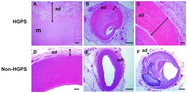
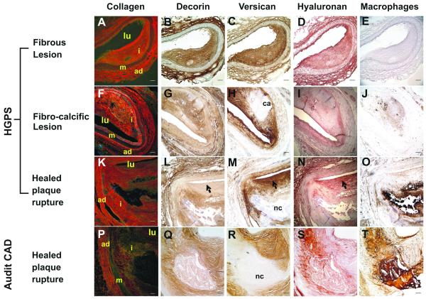
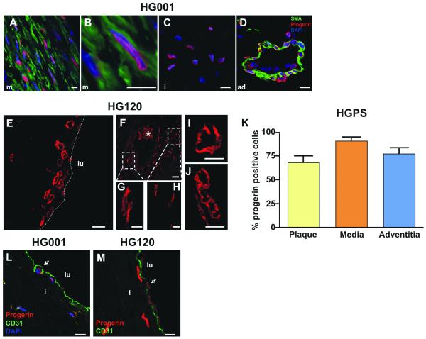
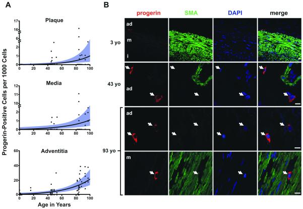
Methods and results: We present structural and immunohistological analysis of cardiovascular tissues from 2 children with HGPS who died of myocardial infarction. Both had features classically associated with the atherosclerosis of aging, as well as arteriolosclerosis of small vessels. In addition, vessels exhibited prominent adventitial fibrosis, a previously undescribed feature of HGPS. Importantly, although progerin was detected at higher rates in the HGPS coronary arteries, it was also present in non-HGPS individuals. Between the ages of 1 month and 97 years, progerin staining increased an average of 3.34% per year (P<0.0001) in coronary arteries.

Conclusions: We find concordance among many aspects of cardiovascular pathology in both HGPS and geriatric patients. HGPS generates a more prominent adventitial fibrosis than typical CVD. Vascular progerin generation in young non-HGPS individuals, which significantly increases throughout life, strongly suggests that progerin has a role in cardiovascular aging of the general population.


Figures

Figure 1
Figure 1. Coronary lesions in HGPS
Movat staining of pathological intimal thickening of A, the LAD and B, RCA of patient HG001. LAD shows a collagen-rich matrix (yellow*) enlarged in the box on the left. Note that the RCA intimal lesion is more cellular (enlarged in the box on the left). C, pathological intimal thickening of the LAD and D, proximal right coronary of patient HG120. The LAD shows a clinically significant stenosis (90%). E, H&E staining of plaques from proximal right coronary of patient HG120 showing a necrotic core (NC); F, cholesterol crystals; G, calcification and H, foam cells. F and G are higher magnification of the regions highlighted in E. (Scale bars: A-E 500 μm; F-H 50 μm).
Figure 2
Figure 2. Fibrosis of the adventitia in HGPS
H&E staining of selected tissues from patient HG001 (A) and HG120 (B-C). A, Aorta with thickened adventitia (arrow). B, Mid-right coronary characterized by an enlarged and highly fibrotic adventitia (arrow). The media is markedly thinned in the area with adventitial fibrosis. C, High-power image of the adventitia (arrow) in B. D, 16-year-old non-HGPS aorta with non-diseased adventitia (arrow). E, 16-year-old non-HGPS LAD, F, 93-year-old LAD with advanced atherosclerosis. The arrow head points to the adventitia. Adventitia: ad, media: m. (Scale bars: A,C-D: 50 μm; B,E-F 500 μm).
Figure 3
Figure 3. ECM deposition in HGPS lesions is similar to that seen in adult CAD
Coronary artery sections were stained for collagen with Picrosirius Red (A, F, K, P), decorin with LF122 (B, G, L, Q), versican with 2B1 (C, H, M, R), hyaluronan (D, I, N, S) and macrophages with CD68 (E, J, O, T). Collagen was imaged using a polarizing filter to distinguish between Type I (orange/red staining) and Type III (green/yellow staining). Black arrow heads refer to patterns of decorin, versican and hyaluronan staining indicative of healed plaque rupture. Vessel lumen: lu, adventitia: ad, media: m, intima: i, calcium deposition: ca, necrotic core: nc. (Scale bars: 100 μm)
Figure 4
Figure 4. Progerin is expressed in coronary vasculature in HGPS
IHC of LAD from patient HG001 (A-D,L) and patient HG120 (E-J,M). A, Anti-progerin-specific Ab staining shows progerin (red) present in medial VSMCs. SMA is stained in green. B, High magnification of a progerin-positive VSMC. C, Progerin-positive cell in the intima and D, in the adventitia. Nuclei are counterstained with DAPI (blue). E-H, Progerin-positive nuclei present in the luminal region of LAD of patient HG120. Boxes in F denote progerin-positive nuclei enlarged in G and H. The dotted line in E represents the luminal border of the artery. Progerin-positive nuclei visible I, in the thickened adventitia and J, in the media. The asterisk denotes autofluorescent red cells. K, quantification of the progerin-positive cells in the plaque, media and adventitia of patient HG001 (LAD). CD31-progerin positive cells denote the presence of EC at the surface of the lumen of the plaque of L, patient HG001 (LAD) and M, patient HG120 (white arrowhead). Nuclei are counter-stained with DAPI (blue). Adventitia: ad, media: m, intima: I, vessel lumen: lu. (Scale bars: A-E,M 10 μm; F 50 μm; G-J 5 μm).
Figure 5
Figure 5. Progerin in coronary arteries of non HGPS subjects with increasing age
A, Progerin-positive cells per 1000 total cells plotted as a function of age in years and the three arterial layers. Lines and bands represent the best fit lines and their 95% confidence intervals as determined by negative binomial general estimating equation, in the plaque, media and adventitia. Samples from the adventitia had significantly higher rates of progerin-positive cells over the entire age range than media (P<0.001) and plaque (P<0.001). The three arterial layers showed significant increases in rate across ages (P<0.0001). B, Representative IHC in a 3-year-old normal control, a 43-year-old CAD patient, and a 93-year-old with advanced-complex plaque. From left to right: progerin (red), SMA (green), DAPI (blue) and merged images. Bottom: example of progerin-positive cell that is not SMA-positive in the media of a 93-year-old with CAD. The arrows indicate the progerin-positive cells. Adventitia: ad, media: m, intima:i. (Scale bars: 10 μm).
Figure 6
Figure 6. Valve and endocardium pathology in HGPS
Valves: A, The mitral valve of patient HG001 is thick and degenerated with visible calcification. Both the fibrosa (F) and the atrialis (A) are expanded. B, the spongiosa (S) is myxomatous and markedly expanded. The boxed area is shown at higher magnification on the right. The arrow points to high ECM content. C, IHC showing expression of progerin (red) in the valves of HG001, DAPI (blue). D, Endocardium of the left ventricle (LV) is thickened compared with another region of the LV (E) with normal endocardium (H&E). Higher magnification is shown on the left corner. IHC: F, the endocardium contains abundant progerin-positive cells (red). DAPI (blue). The arrows in D and F indicate the enlarged endocardium. (Scale bars: A-B,D-E 500 μm; C,F 50 μm).

References

    1. Hennekam RC. Hutchinson-Gilford progeria syndrome: review of the phenotype. Am J Med Genet A. 2006;140:2603–2624. - PubMed
    1. De Sandre-Giovannoli A, Bernard R, Cau P, Navarro C, Amiel J, Boccaccio I, Lyonnet S, Stewart CL, Munnich A, Le Merrer M, Levy N. Lamin a truncation in Hutchinson-Gilford progeria. Science. 2003;300:2055. - PubMed
    1. Eriksson M, Brown WT, Gordon LB, Glynn MW, Singer J, Scott L, Erdos MR, Robbins CM, Moses TY, Berglund P, Dutra A, Pak E, Durkin S, Csoka AB, Boehnke M, Glover TW, Collins FS. Recurrent de novo point mutations in lamin A cause Hutchinson-Gilford progeria syndrome. Nature. 2003;423:293–298. - PMC - PubMed
    1. Shumaker DK, Dechat T, Kohlmaier A, Adam SA, Bozovsky MR, Erdos MR, Eriksson M, Goldman AE, Khuon S, Collins FS, Jenuwein T, Goldman RD. Mutant nuclear lamin A leads to progressive alterations of epigenetic control in premature aging. Proc Natl Acad Sci U S A. 2006;103:8703–8708. - PMC - PubMed
    1. Rusinol AE, Sinensky MS. Farnesylated lamins, progeroid syndromes and farnesyl transferase inhibitors. J Cell Sci. 2006;119:3265–3272. - PubMed

Publication types