. 2010 Oct;6(7):901-11.
doi: 10.4161/auto.6.7.13287. Epub 2010 Oct 2.

Gigantic macroautophagy in programmed nuclear death of Tetrahymena thermophila

Affiliations

Gigantic macroautophagy in programmed nuclear death of Tetrahymena thermophila

Takahiko Akematsu et al. Autophagy. 2010 Oct.

Abstract

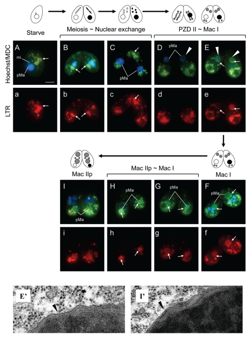
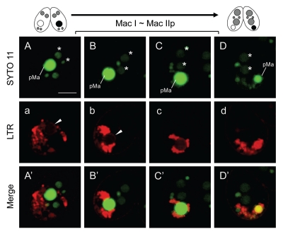
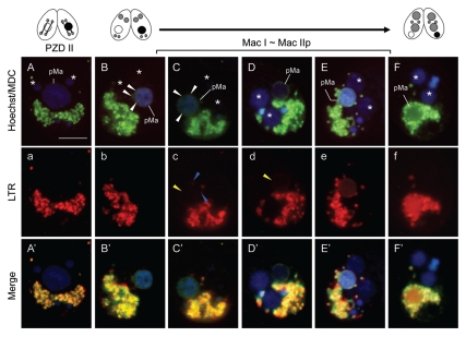
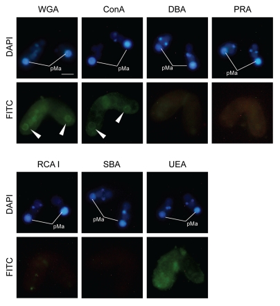
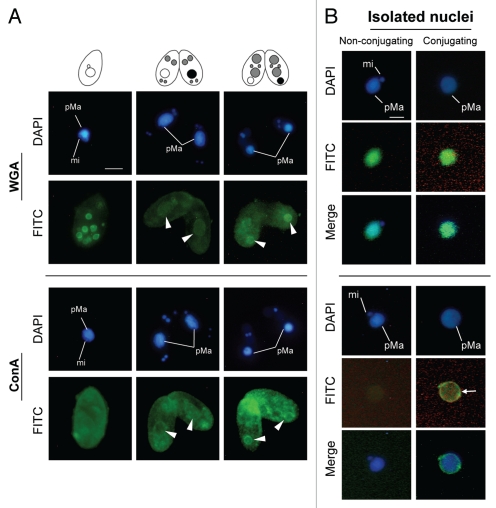
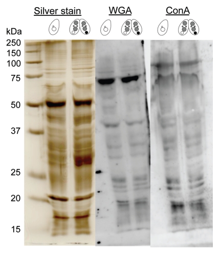
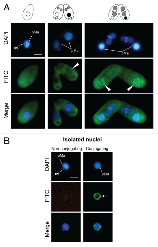
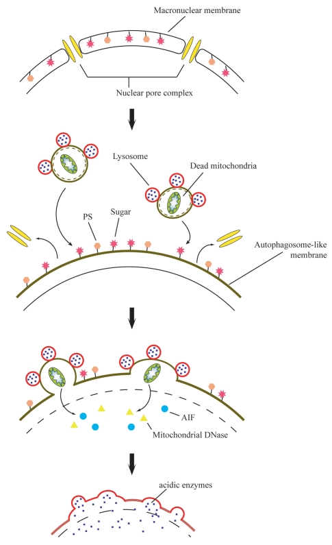
Programmed nuclear death (PND) in Tetrahymena is a unique process during conjugation, in which only the parental macronucleus is degraded and then eliminated from the progeny cytoplasm, but other co-existing nuclei such as new micro- and macronuclei are unaffected. PND through autophagic elimination is expected to be strictly controlled, considering the significant roles in ciliates such as turnover of disused organelles and production of the next generation. Here we demonstrate that PND in Tetrahymena involves peculiar aspects of autophagy, which differ from mammalian or yeast macroautophagy. Drastic change of the parental macronucleus occurs when differentiation of new macronuclei is initiated. Combined use of monodansylcadaverine and a lysosome indicator LysoTracker Red showed that prior to nuclear condensation, the envelope of the parental macronucleus changed its nature as if it is an autophagic membrane, without the accumulation of a pre-autophagosomal structure from the cytoplasm. Subsequently, lysosomes approached only to the parental macronucleus and localized at the envelope until a final resorption stage. In addition, we found that the parental macronucleus exhibits certain sugars and phosphatidylserine on the envelope, which are possible "attack me" signals, that are not found on other types of nuclei. These findings suggest that PND is a highly elaborated process, different from the typical macroautophagy seen in other systems, and is executed through interaction between specific molecular signals on the parental macronuclear envelope and autophagic/lysosomal machineries.


Figures

Figure 1
Figure 1
Autophagic/lysosomal events during PND visualized with a combination of monodansylcadaverine (MDC; upper case) and LysoTracker Red (LTR; lower case). Living cells were stained with MDC and Hoechst 33342 (nuclear stain; upper) and LTR (lower). (A/a) Starved nonconjugating cell. Almost all MDC-sensitive vacuoles co-localize with lysosomes (arrows). (B/b) Meiotic prophase. After initiation of conjugation, these vacuoles accumulate in the posterior region of the cells. (C/c) Nuclear exchange-stage. Many MDC/LTR-staining vacuoles appear in the anterior region of the cells. (D/d and E/e) Transition stage from PZD II to Mac I. Envelope of the parental macronucleus becomes MDC sensitive (arrowheads). (F/f) Mac I-stage cell. Several MDC/LTR-sensitive vacuoles migrate from the posterior region of the cell to the surface of the parental macronucleus (arrows). (G/g and H/h) Transition stage from Mac I to Mac II p. The parental macronucleus translocates to the posterior region of the cell, accompanied by more migration of the vacuoles on their surface (arrows). (I/i) Mac II p-stage. The entire parental macronucleus becomes both MDC- and LTR-sensitive. Transmission electron micrographs of parental macronucleus at two different stages (E′ and I′) are corresponding to (E and I) stages. Black arrowheads in these micrographs indicate the envelope of the parental macronucleus. pMa and mi denote parental macronucleus and micronucleus, respectively. The scale bar in (A) indicates 10 µm.
Figure 2
Figure 2
Confocal imaging of parental macronuclear acidification during Mac I ∼Mac IIp. Living cells in the transition stage from Mac I to Mac IIp were stained with MDC and SYTO 11 nucleic acid stain (upper case; top part) and LTR (lower case; middle part). The lower parts (upper case′) show a merged image. (A/a/A′ and B/b/B′) The envelope of the parental macronucleus shows LTR-positive staining (arrowhead) but the inside is LTR-negative. (C/c/C′) The inside of the parental macronucleus is gradually acidifying by attachment of lysosomes on the envelope. (D/d/D′) The entire parental macronucleus becomes acidic. Arrows in the middle micrographs indicate the envelope of the parental macronucleus. pMa and asterisk (*) denote the parental macronucleus and the new macronucleus, respectively. The scale bar in (A) indicates 10 µm.
Figure 3
Figure 3
Biogenesis of the autophagosomal property on the parental macronuclear envelope. Living cells in the transition stage from PZD II to Mac IIp were stained with MDC and Hoechst 33342 (upper case; top part) and LTR (lower case; middle part). The lower parts show a merged image (upper case′; lower part). (A/a/A′) PZD II-stage cell. (B/b/B′–F/f/F′) Transition stage from Mac I to Mac IIp. Concomitant with parental macronuclear condensation, a green-colored haze of the MDC fluorescence is hanging over the nucleus (white arrowheads in B and C) and then covered the envelope (white arrowhead in D). The envelope was LTR-negative at first (b) and this became slightly LTR-positive after the beginning of the MDC-stainability (yellow arrowheads in c and d) owing to the attachment of several digestive vesicle complexes (blue arrowheads in c). After that, more migration of the digestive vesicle complexes occurred (E and e) and this allowed for acidification of the nucleus (F and f). pMa and asterisk (*) denote the parental macronucleus and the new macronucleus, respectively. The scale bar in (A) indicates 10 µm.
Figure 4
Figure 4
Lys 4 methylation of histone H3 in the new and parental macronuclei during conjugation. (A/A′/A″) Early stage of Mac I. (B/B′/B″) Later stage of Mac I. Lys 4 of H3 in the parental macronucleus (M) is methylated, that in the new macronucleus (A/A′/A″) is not at this stage. (C/C′/C″) Mac IIp. Just after nuclear differentiation, the methyl mark is only detected in the developing macronuclei, while it has disappeared in the degenerating parental macronucleus (dM). Lys 4 of H3 in the micronucleus (m) is not methylated throughout the life cycle. Left, middle and right parts show propidium iodide (upper case; DNA), a-MeH3K4 (upper case′) and a merged image (upper case″), respectively.
Figure 5
Figure 5
Binding of lectin to the parental macronucleus. The Mac IIp-stage cells were fixed and stained with various FITC-labeled lectin probes. WGA: Wheat germ agglutinin. ConA: Concanavalin A. DBA: Dolichos biflorus agglutinin. PRA: Peanut root lectin. RCA I: Ricinus communis agglutinin. SBA: Soybean agglutinin. UEA: Ulex europaeus agglutinin. The FITC fluorescence is concentrated around the parental macronucleus when WGA or ConA are applied (arrow heads). pMa denotes the parental macronucleus. The scale bar in the photograph indicates 10 µm.
Figure 6
Figure 6
Increase of lectin binding in the parental macronucleus. (A) Cells were fixed and stained with FITC-labeled WGA or ConA probes. FITC fluorescence is concentrated around the parental macronucleus at the nuclear degeneration stage (arrowheads). (B) Isolated nuclei were stained with FITC-labeled WGA or ConA probes. The degenerating macronucleus shows an affinity with ConA in the envelope (arrow). pMa and mi denote the parental macronucleus and micronucleus, respectively. The scale bar in (A) indicates 10 µm.
Figure 7
Figure 7
Comparative analysis of nuclear glycoprotein present in starved cells and Mac IIp-stage cells. Nuclei were isolated from starved cells or Mac IIp-stage cells and the lysates were analyzed by lectin-western blotting. The pattern of separated proteins that was visualized by silver staining is also shown. Positions of markers are indicated at the left with molecular masses in kilodaltons (kDa).
Figure 8
Figure 8
Exhibition of an apoptotic molecular marker on the parental macronucleus. (A) The cells were fixed and stained with a FITC-labeled annexin V probe. The FITC fluorescence is concentrated around the parental macronucleus reflecting nuclear degeneration (arrowheads). (B) Isolated nuclei were stained with FITC-labeled annexin V probe. The degenerating macronucleus shows an affinity for annexin V in the envelope (arrow). pMa and mi denote the parental macronucleus and micronucleus, respectively. The scale bar in (A) indicates 10 µm.
Figure 9
Figure 9
Cartoon illustrating a possible sequence of events leading to the unique atophagic/lysosomal nuclear degradation in Tetrahymena. Concomitant with nuclear condensation, alteration of the macronuclear envelope occurs, which is similar to autophagosome formation. Sugars and phosphatidylserine (PS) restricted in the inner leaflet are exposed on their surface at the nuclear alteration stage. These act as an “attack-me” signal, which is responsible for the attraction of the digestive vacuole complexes. The two sorts of digestive vesicles fuse with the nuclear envelope stepwise and release their contents into the nucleus at distinct stages, the initial stage of DNA degradation and the final resorption stage.

References

    1. Mochizuki K, Gorovsky MA. Small RNAs in genome rearrangement in Tetrahymena. Curr Opin Gent Dev. 2004;14:181–187. - PubMed
    1. Davis MC, Ward JG, Herrick G, Allis CD. Programmed nuclear death: apoptotic-like degradation of specific nuclei in conjugating Tetrahymena. Dev Biol. 1992;154:419–432. - PubMed
    1. Mpoke S, Wolfe J. DNA digestion and chromatin condensation during nuclear death in Tetrahymena. Exp Cell Res. 1996;225:357–365. - PubMed
    1. Kobayashi T, Endoh H. Caspase-like activity in programmed nuclear death during conjugation of Tetrahymena thermophila. Cell Death Differ. 2003;10:634–640. - PubMed
    1. Yakisich JS, Kapler GM. The effect of phosphoinositide 3-kinase inhibitors on programmed nuclear degradation in Tetrahymena and fate of surviving nuclei. Cell Deth Differ. 2004;11:1146–1149. - PubMed

Publication types

LinkOut - more resources