. 2010 Oct;28(10):1882-92.
doi: 10.1002/stem.508.

ETV5 regulates sertoli cell chemokines involved in mouse stem/progenitor spermatogonia maintenance

Affiliations
Free PMC article

ETV5 regulates sertoli cell chemokines involved in mouse stem/progenitor spermatogonia maintenance

Liz Simon et al. Stem Cells. 2010 Oct.
Free PMC article

Abstract

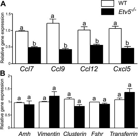
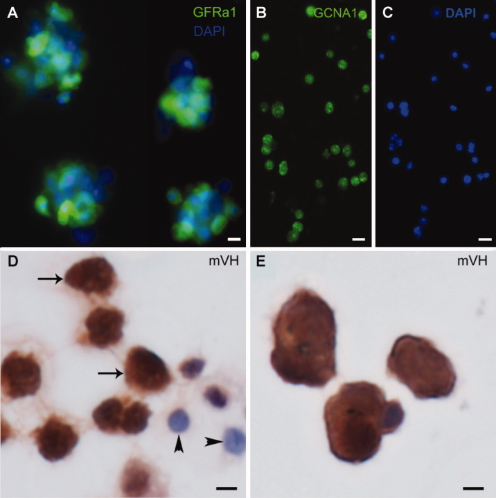
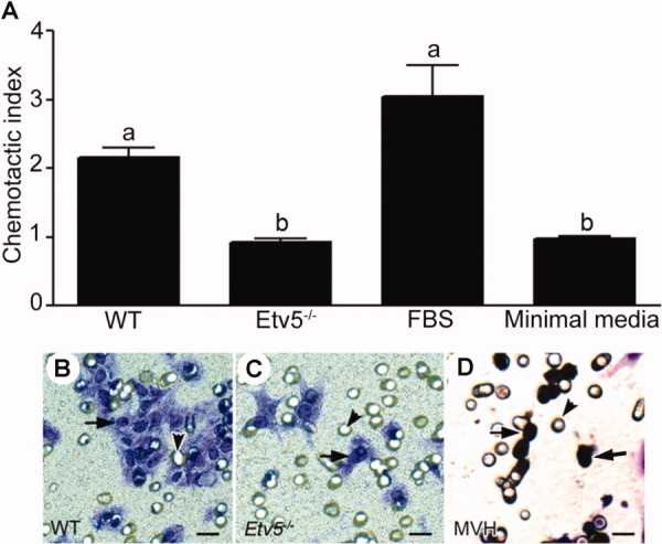
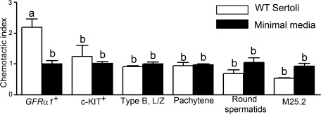
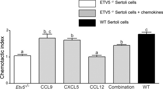
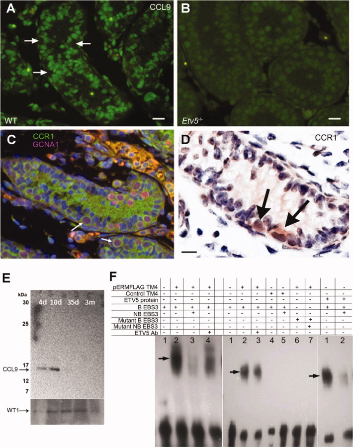
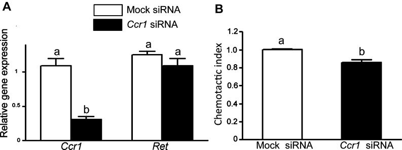
Spermatogonial stem cells are the only stem cells in the body that transmit genetic information to offspring. Although growth factors responsible for self-renewal of these cells are known, the factors and mechanisms that attract and physically maintain these cells within their microenvironment are poorly understood. Mice with targeted disruption of Ets variant gene 5 (Etv5) show total loss of stem/progenitor spermatogonia following the first wave of spermatogenesis, resulting in a Sertoli cell-only phenotype and aspermia. Microarray analysis of primary Sertoli cells from Etv5 knockout (Etv5(-/-)) versus wild-type (WT) mice revealed significant decreases in expression of several chemokines. Chemotaxis assays demonstrated that migration of stem/progenitor spermatogonia toward Etv5(-/-) Sertoli cells was significantly decreased compared to migration toward WT Sertoli cells. Interestingly, differentiating spermatogonia, spermatocytes, and round spermatids were not chemoattracted by WT Sertoli cells, whereas stem/progenitor spermatogonia showed a high and significant chemotactic index. Rescue assays using recombinant chemokines indicated that C-C-motif ligand 9 (CCL9) facilitates Sertoli cell chemoattraction of stem/progenitor spermatogonia, which express C-C-receptor type 1 (CCR1). In addition, there is protein-DNA interaction between ETV5 and Ccl9, suggesting that ETV5 might be a direct regulator of Ccl9 expression. Taken together, our data show for the first time that Sertoli cells are chemoattractive for stem/progenitor spermatogonia, and that production of specific chemokines is regulated by ETV5. Therefore, changes in chemokine production and consequent decreases in chemoattraction by Etv5(-/-) Sertoli cells helps to explain stem/progenitor spermatogonia loss in Etv5(-/-) mice.


Figures

Figure 1
Figure 1
mRNA levels of chemokines are decreased in Etv5-/- Sertoli cells. (A): mRNA levels of Ccl7, Ccl9, Ccl12, and Cxcl5 in freshly isolated Sertoli cells of Etv5-/- mice were decreased 50%–70% compared with age-matched WT mice as determined by real-time polymerase chain reaction. (B): mRNA expression of other Sertoli cell genes. Anti-Mullerian hormone (Amh), vimentin, clusterin, follicle stimulating hormone receptor (Fshr), and transferrin were not altered in Etv5-/- mice. Results are expressed as mean ± SEM (n = 3 for each genotype). Values with different letters were significantly different (p < .01). Abbreviations: ETV5, Ets variant gene; WT, wild type.
Figure 2
Figure 2
Stem/progenitor spermatogonia and other germ cells isolated by STAPUT. Different germ cell populations were isolated from C57Bl/6 mice at specific ages using the STAPUT method. (A): Type A spermatogonia were further subjected to magnetic-activated cell sorting for enrichment in stem/progenitor spermatogonia. About 80% of these cells show intense immunofluorescence for GFRa1, a spermatogonial stem cell marker (green Alexa Fluor 488 staining). Scale bar = 10 µm. (B): These cells also express GCNA1, a germ cell marker (green Alexa Fluor 488 staining). Scale bar = 25 µm. (C): DAPI staining for cells expressing GCNA1. Scale bar = 25 µm. (D): Immunohistochemistry of type B spermatogonia, leptotene/zygotene spermatocytes (arrows) show that they express mVH, an early germ cell marker, and Sertoli cells (arrowheads) do not express the protein. Scale bar = 10 µm. (E): Early pachytene spermatocytes also express mVH. Scale bar = 10 µm. Abbreviations: DAPI, 4',6-diamidino-2-phenylindole; GFRa1, GDNF familiy membrane receptor alpha 1; GCNA1, germ cell nuclear antigen 1; mVH, mouse vasa homolog.
Figure 3
Figure 3
Etv5-/- Sertoli cells have decreased chemoattractive ability. Stem/progenitor spermatogonia were isolated with the STAPUT/magnetic-activated cell sorting method using a GDNF familiy membrane receptor alpha 1 (GFRa1) antibody. They were used for chemotaxis assays toward Sertoli cells using 8-µm-pore transwell units in 24-well plates. The ability of stem/progenitor spermatogonia to migrate toward Sertoli cells was expressed as their chemotactic index. (A): Results indicate that the chemotactic index of stem/progenitor spermatogonia toward WT Sertoli cells was twice more than that toward Etv5-/- Sertoli cells. The chemotactic index toward the positive control (10% FBS) was approximately threefold that of the negative control (minimal media). The results are representative of three independent experiments and are expressed as mean ± SEM. Values with different letters were significantly different (p < .01). (B–D): The panel shows representative images of migrated cells. (B): Stem/progenitor spermatogonia after migration toward WT Sertoli cells and (C) after migration toward Etv5-/- Sertoli cells as a comparison. There was a marked decrease in the number of cells that migrated toward Etv5-/- Sertoli cells. (D): mVH expression of migrated cells showed that more than 95% of the chemoattracted cells were germ cells (arrows). Arrowheads represent the 8 µm pores of the transwell unit. Scale bar = 10 µm. Abbreviations: Etv5, Ets variant gene; FBS, fetal bovine serum; mVH, mouse vasa homolog; WT, wild type.
Figure 4
Figure 4
Chemoattractive ability of WT Sertoli cells is specific for stem/progenitor spermatogonia. Chemotaxis assays were performed to assess if Sertoli cells had a specific effect on GFRa1+ cells compared with cKIT+, type B spermatogonia, leptotene/zygotene spermatocytes, pachytene spermatocytes, or round spermatids. A mesoangioblast stem cell line (M25.2) was also used as a control. The chemotactic index of stem/progenitor spermatogonia (GFRa1+) toward WT Sertoli cells was significantly greater than all other cell types. The chemotactic index of germ cell fractions other than GFRa1+ cell fraction migrating toward WT Sertoli cells was not significantly different from that migrating toward minimal media alone. The results are representative of three independent experiments and are expressed as mean ± SEM. Values with different letters were significantly different (p < .05). Abbreviations: GFRa1, GDNF familiy membrane receptor alpha 1; L/Z, leptotene/zygotene spermatocytes; type B, type B spermatogonia; WT, wild type.
Figure 5
Figure 5
Chemokines increase the chemoattractive ability of Etv5-/- Sertoli cells. Recombinant proteins CCL9, CXCL5, and CCL12 were added individually or in combination along with Etv5-/- Sertoli cells. CCL9 and CXCL5 significantly increased the chemoattractive ability of Etv5-/- Sertoli cells. Addition of CCL9 was able to restore the chemoattractive ability of Etv5-/- Sertoli cells to WT levels. Adding CCL12 did not increase the chemoattraction of these cells. The combination of all three chemokines significantly increased the chemoattractive ability of Etv5-/- Sertoli cells, but did not restore it to WT levels. Results are representative of three independent experiments and are expressed as mean ± SEM. Values with different letters were significantly different (p < .05). Abbreviations: CCL9, C-C-motif ligand 9; CXCL5, C-X-C motif ligand 5; CCL12, C-C-motif ligand 12; Etv5, Ets variant gene; WT, wild type.
Figure 6
Figure 6
Expression of CCL9 in mouse testis. (A): Immunofluorescence of 8-day WT mouse testis showing strong expression of CCL9 in Sertoli cells (green Alexa Fluor 488 staining). Arrows indicate locations of stem/progenitor spermatogonia, which are negative for CCL9. Scale bar = 25 µm. (B): Immunofluorescence of 8-day Etv5-/- mouse testis showing a sharp decrease of CCL9 expression in Sertoli cells. Scale bar = 15 µm. (C): Immunofluorescence of 8-day WT mouse testis showing expression of CCR1, the receptor for CCL9, in stem/progenitor spermatogonia. Type A spermatogonia show double-staining for CCR1 (green Alexa Fluor 488 staining) and GCNA1 (pink Alexa Fluor 594 staining). The green staining in the lumen is background staining. Scale bar = 25 µm. (D): Immunohistochemistry of 6-day WT mouse testis showing expression of CCR1 in stem/progenitor spermatogonia (arrows). Scale bar = 15 µm. (E): Western blot of protein extracts from isolated WT seminiferous tubules of 4-, 10-, 35- and 90-day-old mice with anti-CCL9 and anti-WT1 antibodies, showing that CCl9 expression is restricted to early steps of spermatogenesis. (F): Electrophoretic mobility shift assays to investigate ETV5 binding to its consensus sequence in the Ccl9 promoter. The left panel shows gel shifts using nuclear extracts of TM4 cells after transfection with the pERMFLAG construct (pERMFLAG TM4). 1: biotinylated probe alone (B EBS3); 2: nuclear extract of pERMFLAG TM4 and B EBS3, with apparent gel shift (arrow); 3: nuclear extract of pERMFLAG TM4 with B EBS3 and nonbiotinylated probe (NB EBS3) showing outcompetition; and 4: nuclear extract of pERMFLAG TM4 with B EBS3 and ETV5 antibody (ETV5 Ab) showing a decrease of DNA/protein binding. The middle panel shows gel shifts using again pERMFLAG TM4 nuclear extracts. 1: B EBS3 alone; 2: pERMFLAG TM4 with B EBS3, showing apparent gel shift (arrow); 3: pERMFLAG TM4 with B EBS3 and ETV5 Ab showing decrease of protein-DNA binding; 4: nuclear extract of TM4 cells after transfection with control vector (Control TM4), and incubated with B EBS3; 5: control TM4 with B EBS3 and NB EBS3, 6: pERMFLAG TM4 with mutant B EBS3, and 7: pERMFLAG TM4 with mutant NB EBS3. Gel shifts with NB EBS3 substantially, but not completely outcompeted the protein-DNA complex suggesting the presence of other factors in the complex. There was no shift of protein-DNA complexes with mutant EBS3 sequences or TM4 cells transfected with control vector. The right panel shows gel shifts of purified ETV5 protein using pET vector (ETV5 protein). 1: ETV5 protein with B EBS3, showing gel shift (arrow) and 2: ETV5 protein with B EBS3 and NB EBS3 showing outcompetition. The data confirms a possible ETV5-EBS3 binding. Abbreviations: CCL9, C-C-motif ligand 9; CCR1, C-C-receptor type 1; GCNA, germ cell nuclear antigen 1; Etv5, Ets variant gene; ETV5 protein, Ets variant protein; B EBS3, biotinylated Ets binding site 3; NB EBS3, nonbiotinylated Ets binding site 3; WT, wild type.
Figure 7
Figure 7
RNA interference of Ccr1 in stem/progenitor spermatogonia. (A): RNA interference of Ccr1 in stem/progenitor spermatogonia downregulated the expression of the gene 70% compared with mock siRNA-treated stem/progenitor spermatogonia. Expression of Ret in Ccr1 siRNA transfected stem progenitor/spermatogonia showed that the gene was not affected due to transfection. (B): Chemotaxis assays were performed with stem/progenitor spermatogonia that were transfected with Ccr1 and mock siRNA for 24 hours. The chemotactic index of transfected cells toward wild-type Sertoli cells was 0.86 and 1.002, indicating a 14% reduction in the migration of these cells. Results are representative of three independent biological repeats for RNA interference and two technical repeats for cell migration assays with the stem/progenitor spermatogonia transfected with Ccr1 siRNA. Values are expressed as mean ± SEM. Values with different letters were significantly different (p < .05).

Similar articles

Cited by

References

    1. Orwig KE. Ryu BY. Master SR, et al. Genes involved in post-transcriptional regulation are overrepresented in stem/progenitor spermatogonia of cryptorchid mouse testes. Stem Cells. 2008;26:927–938. - PMC - PubMed
    1. He Z. Jiang J. Kokkinaki M, et al. Nodal signaling via an autocrine pathway promotes proliferation of mouse spermatogonial stem/progenitor cells through Smad2/3 and Oct-4 activation. Stem Cells. 2009;27:2580–2590. - PMC - PubMed
    1. Ahmed EA. de Rooij DG. Staging of mouse seminiferous tubule cross-sections. Methods Mol Biol. 2009;558:263–277. - PubMed
    1. Orth JM. Proliferation of Sertoli cells in fetal and postnatal rats: A quantitative autoradiographic study. Anat Rec. 1982;203:485–492. - PubMed
    1. Hess RA. Cooke PS. Hofmann MC, et al. Mechanistic insights into the regulation of the spermatogonial stem cell niche. Cell Cycle. 2006;5:1164–1170. - PMC - PubMed

Publication types

MeSH terms